
(Credit: © BuyOutFelix03/peopleimages.com - stock.adobe.com)
Menopause Brain Scans Overturn a Long-Standing Assumption
In A Nutshell
- Women transitioning through menopause showed no significant decline in total or cortical brain volume, while women who stayed premenopausal or postmenopausal throughout the study did lose volume.
- Puberty and pregnancy both come with clear, measurable shrinkage in the brain’s outer layer; menopause did not follow that same pattern.
- Deeper brain structures still lost volume during the menopause transition, so the pause applied specifically to the outer layer, not the whole brain.
- The direct comparison between the three menopause groups fell short of statistical significance, so researchers call the pattern suggestive rather than proven.
Menopause doesn’t appear to speed up gray matter loss. In a new brain-scanning study published in the journal Nature Communications, some of the volume decline normally seen in midlife seemed to level off during the transition itself, at least in the brain’s outer layer. That’s the unexpected pattern from a large study tracking more than a thousand women and girls through puberty, pregnancy, or menopause.
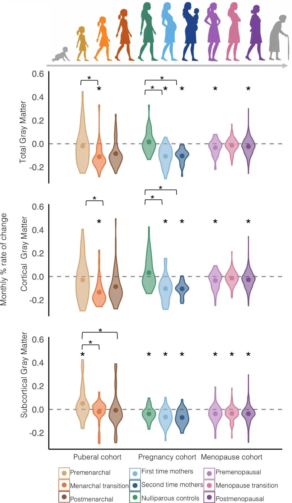
Researchers scanned participants before and after they moved through one of these three hormonal turning points, comparing each group against women and girls who were not actively transitioning. Girls scanned around their first period and women scanned before and after pregnancy both showed clear shrinkage in the cerebral cortex, the brain’s outer layer, matching what smaller studies have hinted at for years. Menopause told a different story. Women moving through the transition showed no real decline in that same outer layer, while women who stayed premenopausal or postmenopausal throughout the study did lose volume there. Deeper brain structures were the exception: those kept shrinking in the transition group too, at roughly the same modest pace seen across the board in midlife. So the pause was specific to the brain’s outer layer, not a blanket freeze on aging.
That pattern complicates a common assumption. Menopause is often described as a period of faster aging or mental fog, and earlier menopause research had reported changes in the hippocampus, a structure tied to memory. Researchers wanted to know whether menopause would follow a pattern similar to puberty and pregnancy, or whether declining hormones might produce something else entirely. The researchers describe menopause as “a qualitatively distinct pattern, defined by the absence rather than acceleration of volumetric change.”
Menopause Brain Aging Pattern Differs Sharply From Puberty and Pregnancy
Few prior studies had compared all three hormonal transitions using identical methods in one project. This team pulled together three groups of participants and ran the same scanning analysis on each.
A group of 142 girls and young women recruited through local schools in the Netherlands made up the puberty cohort, split by whether they had reached their first period yet. A second group of 110 women becoming mothers for the first or second time, plus a childless comparison group, made up the pregnancy cohort. Women drawn from the UK Biobank, a large health database, formed by far the largest group at 843 participants, divided into those who stayed premenopausal, stayed postmenopausal, or moved through the transition.
Every participant received at least two brain scans, spaced months or years apart. Menstrual and menopause status relied on self-reported questions rather than lab testing, a limitation the authors acknowledge.
Brain Scans Show Menopause Volume Loss Levels Off, Not Stops
Girls moving through their first period showed measurable monthly shrinkage in gray matter, a pattern absent in girls who had not yet started menstruating and in girls well past that point. Women becoming mothers for the first or second time showed a similar rate of shrinkage, echoing earlier work from the same research group suggesting pregnancy and the menarche transition may involve comparable brain remodeling. That said, the apparent difference between the transitioning menopause group and the two stable menopause groups fell short of statistical significance when all three were compared directly against each other, so that particular finding reads as suggestive rather than settled.
Region by region, puberty and pregnancy overlapped heavily in areas tied to higher-level thinking and social processing, though they pulled apart in more than half of the 74 brain regions researchers examined. In areas tied to movement and coordination, a step-down pattern emerged: puberty showed the steepest decline, pregnancy came next, and menopause showed the least change of all three. Among first-time mothers, older maternal age was linked to a stronger version of the pregnancy brain-change pattern, though that link did not show up in second-time mothers or in women without children.
Menopause Brain Aging Research Fills a Long-Standing Gap
None of this means menopause is easy or free of symptoms, and researchers are careful to say their scan data cannot show whether this pause is good, neutral, or something else entirely. It does show that the popular story about menopause speeding up brain aging across the board does not match what full-brain scans reveal.
That distinction matters because research on women’s brains has lagged behind for decades, even though women make up half the population. Only a small share of neuroscience research has focused exclusively on female participants, and pregnancy’s effects on the brain became an active research field only about ten years ago. Menopause lagged even further behind that, with earlier work mostly limited to one small brain structure. Drawing on more than a thousand participants, this study pushes back on a long-held assumption about the female brain as reproductive hormones decline. Puberty and pregnancy appear to remodel the brain through rising hormones. Menopause, where hormones fall instead, seems to work differently: rather than accelerating change, it briefly holds the line. The researchers say future studies that measure hormone levels directly, alongside brain scans, could help confirm why.
Disclaimer: This article summarizes findings from a single peer-reviewed study and is not medical advice. The brain-scan data cannot determine whether the pattern described here is beneficial, harmful, or neutral, and it does not address symptoms such as memory problems or brain fog. Readers with questions about menopause and brain health should speak with a qualified healthcare provider.
Paper Notes
Limitations
The authors point out several caveats worth keeping in mind. Menstrual status and menopause status were both determined through single self-reported questions rather than medical testing, meaning some participants labeled as premenopausal may have already been in the early stages of the transition, and the same applies to girls labeled as not yet having started puberty. The three groups were scanned on different MRI machines without cross-cohort calibration, so direct comparisons between raw numbers across groups call for caution, even though the researchers used a subtraction method designed to reduce that issue. Hormone level data wasn’t available across all three groups, so the study could infer hormonal direction from what’s already known about these transitions but couldn’t directly link specific hormone levels to specific brain changes in every cohort. Information on birth control use, socioeconomic status, mental health, and stress levels wasn’t consistently available either. The subcortical (deeper brain structure) findings were limited to the puberty group, since pregnancy and menopause both showed general declines in that area that made region-specific patterns harder to detect. Finally, the study didn’t include men as a comparison group, a deliberate choice since puberty, pregnancy, and menopause are tied to female biology and comparing across sexes would have muddied the specific effects being studied.
Funding and Disclosures
The project was supported by an Innovational Research Incentives Scheme grant (Veni, 451-14-036) from the Netherlands Organization for Scientific Research, a grant from the Leiden University Fund/Elise Mathilde Fund, and a NARSAD grant from the Brain and Behaviour Research Foundation in the United States, all awarded to Elseline Hoekzema, who is currently supported by an ERC Starting Grant from the European Research Council. The authors reported no competing interests.
Publication Details
The study, titled “Puberty, pregnancy, and menopause show shared and distinct structural changes across the lifespan,” was authored by Sophie R. van ‘t Hof, Marieke G.N. Bos, Milou Straathof, Eveline A. Crone, and Elseline A. Hoekzema. It was published in Nature Communications (2026, volume 17, article 9360). The DOI is 10.1038/s41467-026-76755-2.


