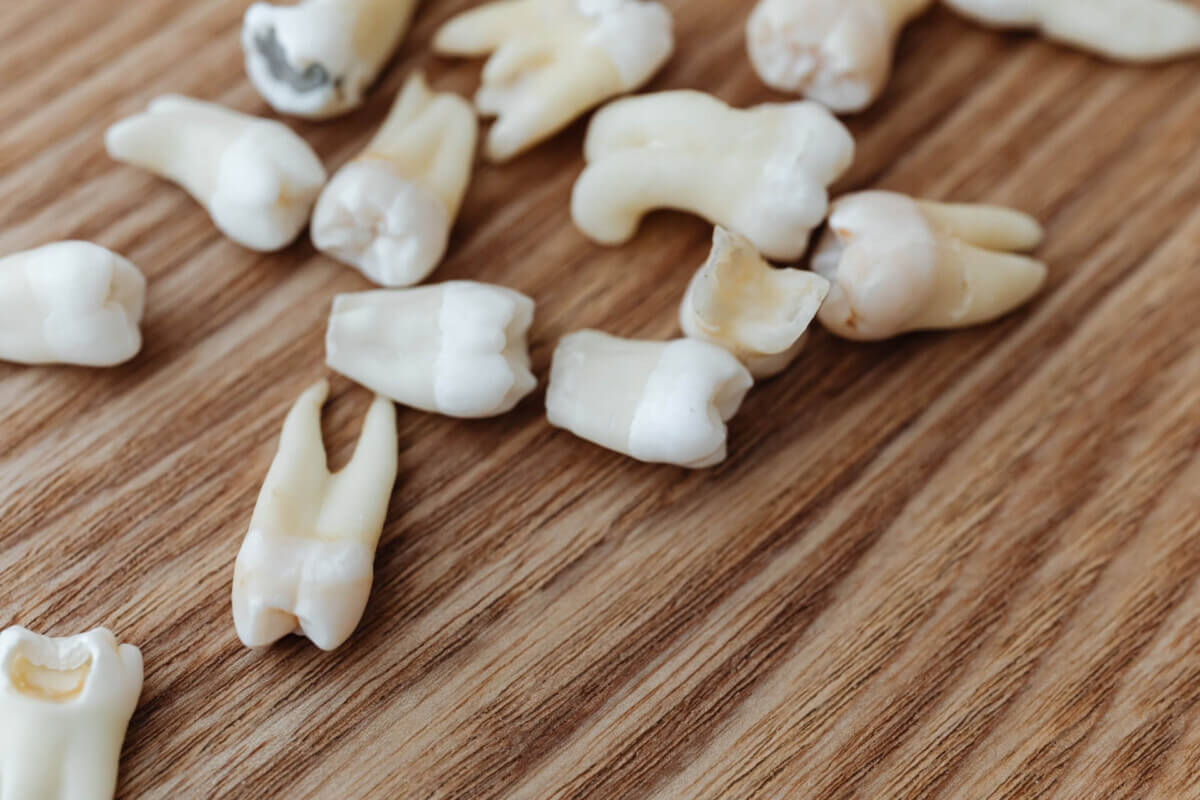
(Credit: Karolina Grabowska from Pexels)
Despite a healthy diet, mice missing teeth showed notable memory loss
In A Nutshell
- Memory declined after tooth loss in mice. Aging mice that had their upper molars extracted on both sides performed worse on memory tests six months later compared to mice that kept their teeth
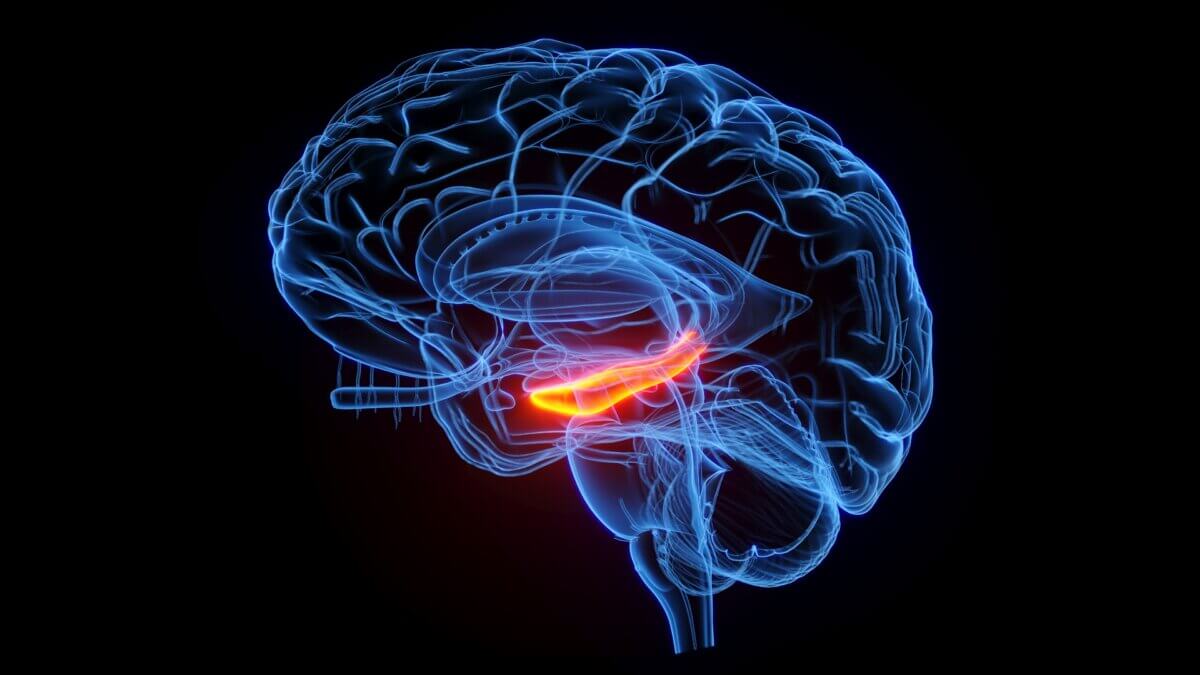
- Brain tissue showed stress markers in memory centers. The hippocampus, which handles memory formation, displayed increased inflammation markers and fewer cells marked as neurons in tooth loss groups.
- Diet alone didn’t explain the brain changes. Mice missing teeth showed these brain alterations whether they ate protein-rich or protein-poor diets, suggesting tooth loss affects the brain through a direct pathway rather than just through nutrition problems.
- Human relevance remains unknown. This study tested mice only and did not measure whether similar processes occur in human brains, though the findings could point researchers toward new questions about oral health and aging.
Losing several teeth changes more than just how well someone can chew a steak. A study from Japan suggests something more concerning happens. After their teeth were removed, studied animals started having memory problems. Their brains showed signs of stress and stress-related changes in the regions that handle memory and learning. What’s more, this happened even when the mice ate a normal-protein diet, suggesting the tooth loss itself, not just poor nutrition, might be affecting the brain.
Scientists at Hiroshima University tracked aging mice for six months after pulling their upper molars on both sides. They wanted to know if tooth loss leads to brain problems because people can’t eat well afterward, or if something else is going on. The answer surprised them. Mice that lost teeth performed worse on memory tests whether they ate normal or low-protein diets. When researchers examined brain tissue, they found higher levels of molecules linked to cell death, increased signs of inflammation, and fewer cells marked as neurons in key memory regions.
These are mice, not people. But the findings, published in Archives of Oral Biology, hint at an effect not explained by diet alone between missing teeth and brain changes that goes beyond just eating less protein.
How Tooth Loss Affects Memory and Cognitive Function
Researchers divided aging mice into four groups at three months old. Some had their upper molars on both sides extracted while others kept all their teeth. Half of each group received normal protein levels in their food, while the other half ate diets containing 50 percent less protein. This setup was designed to approximate what can happen for some elderly people after losing teeth: they avoid meat, fish, and eggs because chewing hurts.
Six months later, the team tested memory using a Barnes maze, a circular platform with 20 holes around its edge. Just one hole led to an escape box. Mice with intact memories learned quickly which hole offered escape. Mice with memory problems took longer to find the correct hole, often following more erratic paths.
The results were clear: mice that lost their teeth performed worse on memory tests than mice with full sets of teeth. Among mice eating less protein, this difference appeared larger, though the study notes its sample size may have been too small to detect whether diet and tooth loss interact statistically. Body weight remained steady across all groups, ruling out starvation or general poor health as explanations.
Most importantly, tooth loss drove the memory problems, whether mice ate normal-protein diets or protein-poor diets. The missing teeth themselves, not dietary protein levels, predicted which mice would struggle to remember.
Brain Cell Death Linked to Tooth Loss
The memory tests revealed problems, but brain tissue analysis showed why. Scientists examined the hippocampus, where the brain forms and stores memories. They measured gene activity for two molecules called Bax and Bcl-2 that regulate cell death pathways. When Bax gene expression rises compared to Bcl-2, it is commonly used as a marker consistent with apoptosis-related signaling, a programmed cell death process.
Mice missing teeth showed higher Bax-to-Bcl-2 ratios in their hippocampi compared to mice with intact teeth. Protein intake made no difference. Whether mice consumed normal or reduced protein, tooth loss was associated with higher levels of this apoptosis-related marker.
Additional analysis revealed markers of inflammation and fewer NeuN-positive cells in specific memory regions. The CA1 region, which helps form new memories and recall old ones, displayed high levels of GFAP and Iba-1, proteins that signal brain inflammation and stress. The same region contained fewer NeuN-positive cells, a marker consistent with reduced neuron presence in the sampled tissue.
The dentate gyrus, another memory region, showed similar patterns: markers of increased inflammation and fewer NeuN-positive cells. The CA3 region showed less change, though low protein did reduce neuron-related cell markers there. Across the hippocampal regions they examined, tooth loss had the dominant association with these brain changes while diet played a smaller role.
The Connection Between Missing Teeth and Brain Health
The relationship between oral health and brain function has interested researchers for years, though the mechanisms remain unclear.
The paper discusses several possibilities. Gum disease, which often precedes tooth loss, involves bacteria and inflammatory processes. Inflammatory signals could potentially affect blood vessels or other brain tissue. Another theory involves sensory input: teeth connect to the brain through the trigeminal nerve, one of the largest nerves in the head. Chewing sends information through this nerve to brain regions handling attention, learning, and memory. Losing teeth disrupts these signals in mice, which might affect brain activity.
This mouse study supports the idea of an effect not solely explained by nutrition. Mice fed a normal-protein diet still showed markers of brain inflammation and reduced NeuN-positive cells after tooth extraction. The tooth loss itself appears associated with brain changes in mice, not just secondary effects like malnutrition.
The research team used aging mice (SAMP8 strain) that naturally develop age-related problems including memory decline, making them useful for studying tooth loss effects in the context of aging.
Study Limitations and What They Mean
Mouse brains differ from human brains, so these findings need confirmation in people before drawing conclusions about tooth loss and dementia in humans. Mice received standardized diets while human nutrition varies widely. The six-month observation period in these aging mice may not capture all relevant time-course effects.
Reducing dietary protein meant increasing carbohydrates to maintain total calories. Higher carbohydrate intake could have influenced results, though this doesn’t change the main finding about tooth loss associations. Sample sizes ranged from seven to nine mice per group; larger studies would provide more confidence in the results, particularly regarding potential interactions between tooth loss and diet. Only male mice were tested, so whether females show the same patterns remains unknown.
Protecting Your Teeth May Protect Your Memory
In these mice, tooth loss was associated with worse memory performance, increased markers of apoptosis-related signaling, heightened neuroinflammation indicators, and fewer NeuN-positive cells in key memory regions. The tooth loss associations and the low-protein diet effects appeared to work through separate mechanisms rather than combining to amplify each other, though the authors note the study may have lacked sufficient statistical power to detect interactions confidently.
Whether similar processes occur in humans requires further study. The mouse findings suggest tooth loss could directly affect brain biology rather than working only through nutritional changes, but proving this in humans needs additional research. If the connection holds, preventing tooth loss might become one strategy for supporting cognitive health during aging.
One open question involves whether replacing missing teeth might help. The study did not test dental implants or dentures, so whether restoring chewing ability after tooth loss could affect brain markers or cognitive function remains unknown. Future research testing this possibility could determine if tooth replacement offers benefits beyond improved nutrition.
Understanding connections between oral health and brain function continues to develop as a research area. The mechanisms linking tooth loss to brain changes in mice could eventually help identify new approaches for supporting healthy aging.
Disclaimer: This article reports on an animal study conducted in mice. The research did not test humans, and whether similar biological processes occur in people remains unknown. The findings represent associations observed in a laboratory setting and should not be interpreted as medical advice. Readers with concerns about tooth loss or cognitive health should consult healthcare professionals.
Paper Notes
Limitations
This study used SAMP8 mice, an accelerated aging model, and findings may not fully translate to humans given differences in biological pathways between species. The sample sizes were small (7-9 mice per group), limiting statistical power for detecting interaction effects. The observation period was six months, which may not capture longer-term effects. The low-protein diet included compensatory increases in nitrogen-free extract (carbohydrates), making it difficult to separate protein restriction effects from relative increases in carbohydrate intake. Only male mice were studied, so results may not apply to females. The study could not completely separate the effects of tooth loss from the effects of altered chewing mechanics.
Funding and Disclosures
This study received funding from the Japan Society for the Promotion of Science (JSPS) KAKENHI. Grant numbers: 20K10035 (H. Oue), 22K17114 (M. Yokoi), and 20K100725 (T. Kubo). The authors declared no competing financial interests or personal relationships that could have influenced the work reported in this paper.
Publication Details
Authors: Rie Hatakeyama, Hiroshi Oue (corresponding author), Miyuki Yokoi, Eri Ishida, Takayasu Kubo, Kazuhiro Tsuga
Affiliations: Department of Advanced Prosthodontics, Hiroshima University Graduate School of Biomedical and Health Sciences, Hiroshima, Japan; Department of Dentistry & Oral-Maxillofacial Surgery, Fujita Health University, Aichi, Japan
Journal: Archives of Oral Biology, Volume 180, December 2025, Article 106421 | DOI: 10.1016/j.archoralbio.2025.106421 | Published online: October 11, 2025
| Article type: Open access under CC BY license
Work was conducted using facilities at the Central Laboratory of School of Dentistry at Hiroshima University. English language editing assistance was provided by Editage. All animal procedures complied with the Japan Animal Protection Act and were approved by the Institutional Research Facilities Committee (approval number A20-129).


