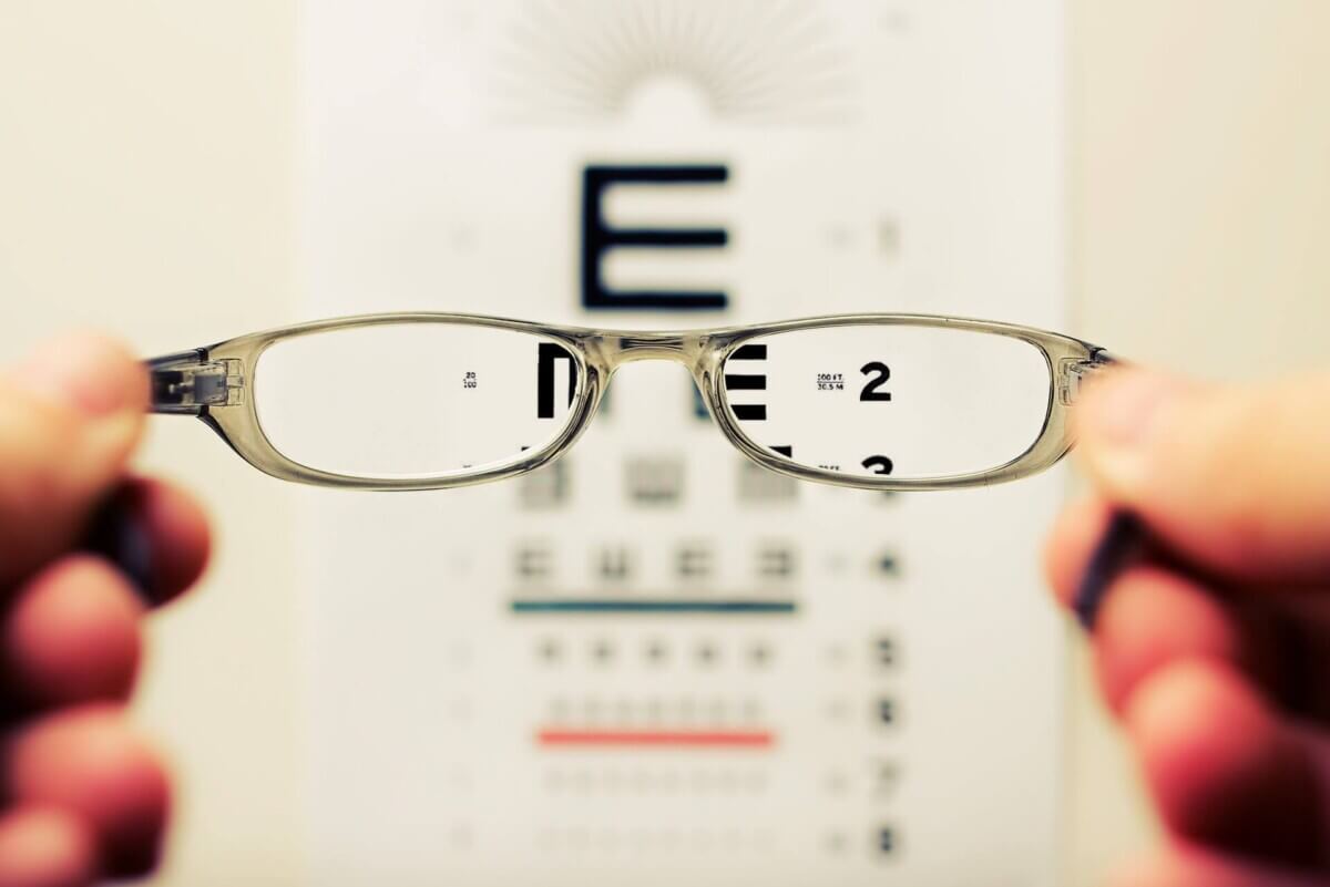
(Photo by David Travis on Unsplash)
Better Glasses Could Be Unlocking Vision That Was Already There, Study Suggests
In A Nutshell
- The findings could help engineers design better visual prosthetics and hold relevance for the roughly 2.2 billion people worldwide living with some form of vision impairment.
- Scientists have found the first direct evidence that most light-sensing cells in the eye, about 76%, each send their signal through a single dedicated connection to the brain.
- Researchers used a specialized imaging tool to correct the eye’s natural blur in real time, revealing brain cells operating at the physical limit of what cone spacing allows.
- If the eye-to-brain wiring has less of a bottleneck than once thought, the quality of a person’s cornea and lens may play a bigger role in sharp vision than previously understood.
At the back of each human eye sits a tiny patch of tissue, smaller than a pinhead, packed with the densest cluster of light-detecting cells in the body. This region, called the fovea, is what lets people read fine print, recognize faces, and thread a needle. Scientists have long believed many of these cells, called cone photoreceptors, send their signal through a dedicated, one-to-one connection to a relay station deep in the brain. The wiring said it should work that way. Actual proof was another matter.
Now researchers from the University of Alabama at Birmingham and the University of California, Berkeley have finally delivered proof. In a study published in Nature Communications, they used a specialized imaging instrument to trace brain-cell activity back to individual cones in the retinas of macaque monkeys. What they found: 76% of the brain cells they studied were each receiving signals from exactly one cone.
That result has a direct consequence. If the wiring from eye to brain has less of a bottleneck than scientists once thought, then the clarity of a person’s optics (the cornea and lens) plays a bigger role in sharpness than previously understood. Better glasses or corrective surgery may be unlocking capacity the visual system already has, rather than compensating for wiring shortfalls.
How the Team Captured the Eye’s Private Line to the Brain
Proving this one-to-one connection had stumped researchers for years because of a stubborn catch-22. To test whether a brain cell responds to a single cone, scientists need to project tiny patterns of light onto the retina with pinpoint accuracy. But the eye’s cornea and lens naturally blur incoming light, smearing fine detail across multiple cones before it even reaches the photoreceptors. It’s like trying to test a camera sensor by shooting through a foggy window.
To get around this, the team used an instrument called an adaptive optics scanning laser ophthalmoscope. It measures the optical imperfections in a living eye and corrects for them using a flexible mirror that reshapes itself 15 times per second to cancel out distortions. Light projected onto the retina then arrives with near-perfect clarity.
With that correction in place, researchers imaged individual cones in three male macaque monkeys while simultaneously projecting flickering red and green light patterns onto those same cells and recording from brain cells in the relay station. Each pattern consisted of tiny squares, each smaller than a single cone, flickering randomly 30 times per second. By tracking which patterns triggered each brain cell to fire, the team mapped exactly which cone was driving it.

Mapping the Brain’s Response Back to Individual Cones
When those maps were overlaid onto actual images of the cone mosaic, the response centers of most brain cells lined up closely, often matching individual cones. Average response-center diameter came out to about 4.2 micrometers, roughly the width of one cone.
To rule out measurement error, the team ran computer simulations modeling how light enters and gets absorbed by cones, then compared predictions for one, two, or three cones feeding a single brain cell against their actual data. Simulation confirmed that 26 of the 34 brain cells studied, or 76%, were most consistent with single-cone input. Six appeared to draw from two cones; two from three.
A secondary test using traditional stripe patterns found these cells responded to visual detail at about 22 cycles per degree of visual angle, roughly four times higher than previous studies had recorded without adaptive optics correction, a gap that shows how much the eye’s natural blur had been masking in earlier work.
Why Earlier Studies Missed This
Earlier attempts typically used stripe patterns or small spots of light, which tend to activate not just the target cone but neighboring ones through side-connections within the retina. Random flickering patterns don’t engage those surrounding circuits, letting the core one-to-one connection come through clearly.
Color-splitting also had to be controlled for, since different wavelengths land on slightly different retinal spots and those offsets vary from eye to eye. Even the animals’ heartbeats posed a problem: pulse-driven eye movements, however tiny, could smear light across multiple cones. Real-time image stabilization, locking the projected patterns to the retina’s actual position 30 times per second, solved that.
What This Means for Everyday Vision and Future Technology
For anyone who has ever squinted at a street sign or struggled to read fine print, this result carries an unusually direct message. There may be less of a bottleneck in the wiring than scientists once thought. Every bit of uncorrected nearsightedness, every slight corneal irregularity, every trace of age-related lens cloudiness can limit how much of that built-in capacity is actually used.
These findings also matter for visual prosthetics, devices designed to restore sight. Knowing the brain’s primary visual pathway is calibrated for single-cone-resolution input gives engineers a more accurate target for what these devices need to deliver.
For the roughly 2.2 billion people worldwide living with some form of vision impairment, the picture is equal parts sobering and encouraging. Extraordinary visual sharpness may already be largely built in, hard-wired from cone to brain. What limits many people isn’t the wiring itself, but often the optics in front of it.
Paper Notes
Limitations
Several limitations are worth noting. Small differences in response-area sizes between red and green light channels in brain cells that responded to both colors could have stemmed from focus differences between the two channels, from color-splitting that was not precisely corrected for each animal, or from larger errors in correction for green light compared to red. In cases where response-area outlines did not fall exactly on the center of a cone, this could have been due to noise in the color-splitting measurements or to undetected shifts in eye position. Notably, four of the six cases classified as two-cone response centers were located less than one degree from the fovea, where the simulations converged and stimulus pixel size was most likely to extend beyond one cone, making it harder to distinguish one-cone from two-cone inputs. The light-capture simulations likely overestimated contributions from additional cones due to various noise sources. All recordings were made from three male macaque monkeys under anesthesia with neuromuscular blockade, which may limit direct generalization to unanesthetized humans.
Funding and Disclosures
This work was supported by National Eye Institute grants R01-EY023581, R01-EY036825, R01-EY023591, R01-EY023603, and R01-EY023603-S1; an Air Force Office of Scientific Research grant (FA9550-21-1-0230); a German Research Foundation grant (TE 1182/1-1); the Eyesight Foundation of Alabama; and a National Eye Institute Core grant (P30-EY003039) to the University of Alabama at Birmingham. Austin Roorda and Pavan Tiruveedhula are co-inventors on patents assigned to the University of California and licensed to C.Light Technologies. No other competing interests were declared.
Publication Details
Title: Physiological basis of resolution acuity in vision | Authors: Keaton M. Ramsey, Philipp Tellers, Alexander Meadway, Pavan Tiruveedhula, Austin Roorda, and Lawrence C. Sincich. Ramsey and Tellers contributed equally. | Affiliations: Neuroengineering PhD Program, School of Engineering and School of Medicine, University of Alabama at Birmingham; Department of Optometry and Vision Science, School of Optometry, University of Alabama at Birmingham; Herbert Wertheim School of Optometry and Vision Science, University of California, Berkeley. | Journal: Nature Communications (2026) 17:2467 | DOI: https://doi.org/10.1038/s41467-026-68851-0 | Received: May 12, 2025 | Accepted: January 19, 2026 | Published online: February 7, 2026 | Corresponding author: Lawrence C. Sincich


