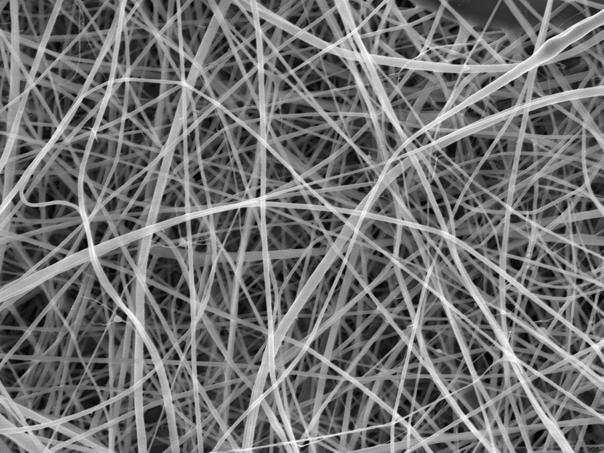
The team used a scanning electron microscope, scanning the mat with a focused beam of electrons and creating an image based on the pattern of electrons that are deflected or knocked-off. Each individual strand is too narrow to be clearly captured by any form of visible light camera or microscope. (Credit: Beatrice Britton / Adam Clancy)
LONDON — What do you get when you cross a pasta maker with a science lab? The world’s thinnest spaghetti, apparently. But while this ultra-thin creation might sound like a culinary achievement, its real potential lies far from the kitchen, in realms ranging from wound healing to tissue regeneration.
Made from ordinary white flour, these incredibly thin fibers, measure just 372 nanometers in diameter — about 200 times thinner than a human hair. To put this in greater perspective, these strands are even narrower than some wavelengths of visible light. The creation, described in the journal Nanoscale Advances, represents a significant advance in the development of sustainable nanomaterials.
While these ultra-thin “nanopasta” strands might sound like an experiment in miniature cooking, they weren’t created for consumption. Rather, they demonstrate a more environmentally friendly approach to creating nanofibers, which have numerous applications in medicine and industry. Traditional methods of creating starch nanofibers require extracting and purifying starch from plant cells, a process that consumes significant amounts of energy and water. This new approach, using ordinary flour directly, offers a simpler and potentially more sustainable alternative.
“To make spaghetti, you push a mixture of water and flour through metal holes. In our study, we did the same except we pulled our flour mixture through with an electrical charge. It’s literally spaghetti but much smaller,” explains co-author Dr. Adam Clancy from UCL Chemistry, in a statement.
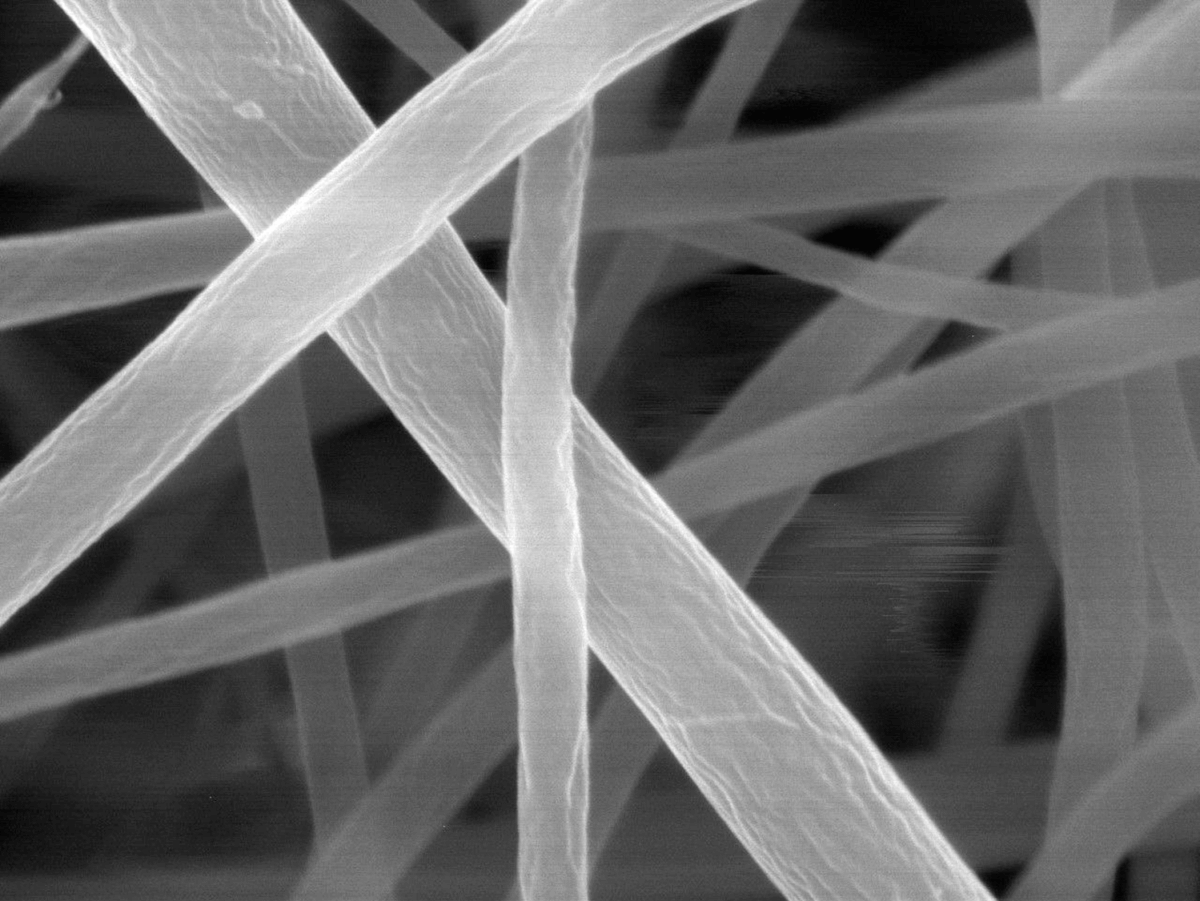
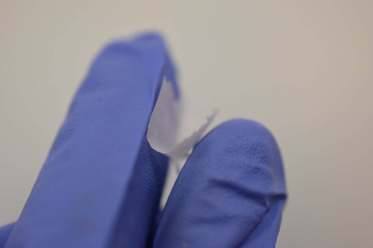
To appreciate just how thin these fibers are, consider that the next thinnest known pasta, called su filindeu (“threads of God”), handmade in Nuoro, Sardinia, is approximately 400 microns wide — about 1,000 times thicker than these new nanofibers. The resulting material forms a mat about two centimeters across, visible to the naked eye, though individual strands are so thin they can only be measured using a scanning electron microscope.
The research team, based in the United Kingdom, developed their process using plain white flour purchased from a Marks & Spencer supermarket. This ordinary flour, containing a precise mixture of carbohydrates (72.8%), protein (9.9%), fiber (2.6%), fat (0.7%), and water (14.7%), proved to be an effective starting material for creating uniform nanofibers.
The process employs a technique called electrospinning, which uses electrical forces to create extremely thin fibers. The researchers discovered that the key to success lay in carefully preparing the flour solution. They mixed the flour with formic acid rather than water, as formic acid breaks down the complex spiral structures (helices) that make up starch molecules. This breakdown is similar to what happens when pasta is cooked, making it digestible, but in this case, it allows the material to form incredibly thin fibers.
Creating these nanofibers from flour proved more challenging than working with pure starch due to the presence of proteins and cellulose, which affect the solution’s viscosity. The researchers found that preparing a 17% flour solution in formic acid, carefully warmed for several hours before cooling, produced the best results. The solution is then pulled through a thin needle by an electric field, with the formic acid evaporating as the fiber travels through the air to a collection plate.
“Starch is a promising material to use as it is abundant and renewable – it is the second largest source of biomass on Earth, behind cellulose – and it is biodegradable, meaning it can be broken down in the body,” Dr. Clancy notes. This biodegradability makes these nanofibers particularly interesting for medical applications.
Professor Gareth Williams from UCL School of Pharmacy explains that such nanofibers show promise for wound dressings due to their porosity, allowing water and moisture to pass while keeping bacteria out. They might also serve as scaffolds for tissue regeneration, mimicking the natural extracellular matrix that supports cell growth.
While these nano-scale spaghetti strands won’t be appearing on dinner plates anytime soon — Professor Williams humorously notes that “it would overcook in less than a second, before you could take it out of the pan” — they represent an important step forward in sustainable materials science. The ability to create functional nanomaterials from common flour opens new possibilities for environmentally friendly medical and industrial applications.
Paper Summary
Methodology
The research team utilized an electrospinning process, which operates like a high-tech version of making pasta. They created a solution using ordinary white flour and formic acid, carefully controlling the temperature and mixing time. The solution was then loaded into a syringe fitted with a thin needle.
When an electric charge was applied, it pulled the solution through the needle, creating extremely thin fibers that collected on a metal plate covered with baking paper. The process required precise control of various parameters, including the distance between the needle and collector (8 cm), flow rate (0.25 mL per hour), and voltage (19-21 kV) for optimal results.
Key Results
The team successfully created uniform nanofibers measuring 372 nanometers in diameter, forming a mat approximately 2 cm across. These fibers showed initial water resistance while remaining biodegradable. The researchers discovered that the protein content in flour actually improved fiber formation compared to pure starch, though this made the process more challenging due to increased viscosity. The study demonstrated that complex natural materials like flour can be successfully transformed into nanoscale fibers without requiring extensive purification.
Study Limitations
Several practical challenges emerged during the research. The process required careful control of temperature and mixing time to achieve the right consistency. The presence of proteins and cellulose in flour made the solution more viscous and harder to work with than pure starch. The resulting films showed some structural limitations, including crazing (cracking) upon drying. Additionally, while visible as a mat, individual fibers were so thin they required specialized electron microscopy for measurement and analysis.
Discussion & Takeaways
This research demonstrates a potentially more sustainable approach to creating nanofibers, bypassing the energy-intensive process of starch purification. The successful use of ordinary flour suggests possibilities for more environmentally friendly production of medical and industrial materials. While not intended for consumption, these ultra-thin fibers could find applications in wound dressings, tissue engineering scaffolds, and other biomedical uses due to their porosity and biodegradability.
Funding & Disclosures
The research was conducted at University College London (UCL), with materials sourced from Sigma-Aldrich and Marks & Spencer. The initial work was performed by Beatrice Britton as part of her master’s degree in chemistry at UCL, demonstrating the educational and academic nature of this research project.


