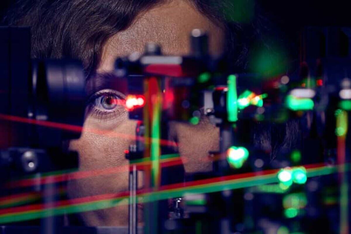
First author Jenny Witten in front of the micro-psychophysical setup. (Credit: MIB-Center at the UKB / Volker Lannert)
BONN, Germany — Even when you think your eyes are perfectly still while reading these words, they’re actually making tiny, unconscious movements. As it turns out, these seemingly random twitches serve a critical purpose: they help you see the world more clearly, according to new research from Germany.
The study, published in the journal eLife, reveals that these microscopic eye movements work in perfect harmony with the arrangement of light-sensing cells in our eyes, automatically adjusting to help us achieve the sharpest possible vision.
“Unlike a camera, our eyes are constantly and unconsciously in motion,” explains Dr. Wolf Harmening, who leads the AOVision laboratory at the University Hospital Bonn’s Department of Ophthalmology, in a media release.
These minuscule movements continue even when we’re trying our hardest to stare at a fixed point.
How do our eyes create sharp vision?
At the center of each eye lies a tiny region called the fovea – a densely packed area of color-sensing cells known as cones. To put its size in perspective, this crucial region is about 200 times smaller than a quarter coin, yet it contains an astounding 200,000 cone cells per square millimeter. These cells are responsible for our ability to see fine details and vivid colors.
Unlike the uniform grid of pixels in your smartphone’s camera, however, these cone cells aren’t evenly distributed. Each person has their own unique pattern of cone density in their fovea, like a fingerprint for vision. What makes this new research particularly fascinating is how our unconscious eye movements – specifically, a type called “drift” – work in concert with this irregular arrangement of cells.
The research team used a specialized instrument called an Adaptive Optics Scanning Light Ophthalmoscope – the only one of its kind in Germany – to conduct their investigation. This high-tech device allowed them to observe both the precise arrangement of cone cells in participants’ eyes and track their minute eye movements simultaneously.
The team studied 16 healthy volunteers, measuring their visual acuity during challenging vision tests while recording exactly how their eyes moved, and which photoreceptor cells were being used. What they found was remarkable: people could actually see finer details than what should have been possible based on the density of their cone cells alone.
The secret is in those tiny, drifting eye movements. Lead author Jenny Witten, a doctoral student at the University of Bonn, found that these movements weren’t random at all.
“The drift movements repeatedly brought visual stimuli into the region where cone density was highest,” explains Witten.
In other words, your eyes automatically drift in patterns that maximize the use of your highest-density cone regions, enhancing your ability to see detail.
The future of better vision
This discovery isn’t just interesting for scientists and eye doctors – it could have important practical applications. Dr. Harmening’s team suggests that understanding how these unconscious eye movements optimize vision could lead to better treatments for various eye disorders and improvements in artificial vision technologies, such as retinal implants.
The next time you find yourself marveling at your ability to read tiny print or appreciate the intricate details in a painting, remember: it’s not just your eyes that are doing the work, but also those constant, invisible movements that help you see the world in crystal clarity.
Paper Summary
Methodology
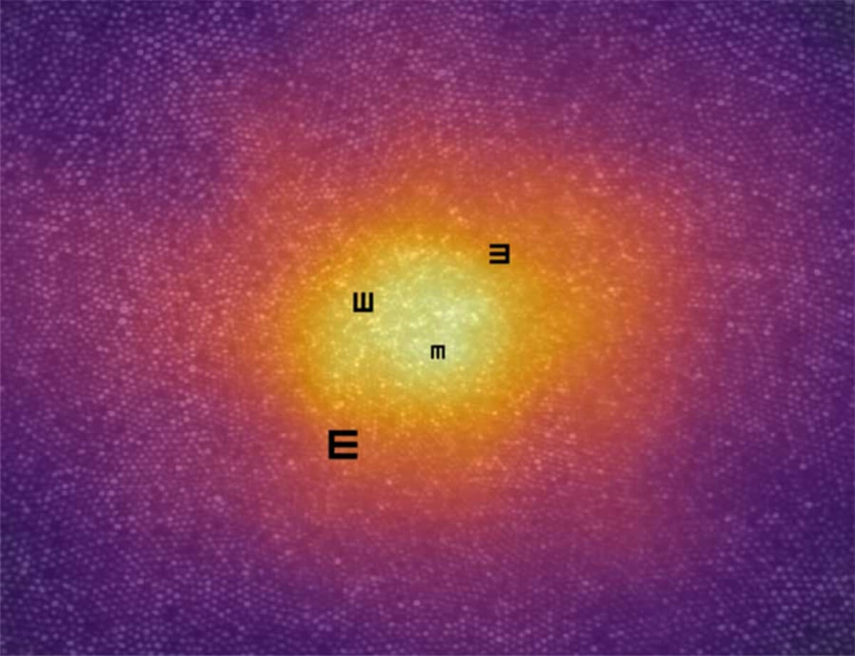
This study explored how tiny movements of our eyes, called “fixational drift,” influence how clearly we see very fine details. The researchers used special high-resolution imaging equipment to look closely at the center of participants’ retinas (the area responsible for our sharpest vision). During the experiment, participants were asked to identify the orientation of a small letter “E” on a screen.
While they looked at the letter, the equipment recorded their eye movements and how different parts of the retina were involved in seeing the image. By analyzing these recordings, the researchers examined how eye movements could help us see even finer details than previously thought possible.
Key Results
The study found that small, controlled eye movements (fixational drift) helped participants see details smaller than a single photoreceptor cell (the tiny light-sensitive cells in the eye). The results showed that these eye movements are not random but are directed toward areas with the highest concentration of these photoreceptor cells. This adaptation allowed participants to see finer details than if their eyes remained still, suggesting that the human visual system uses both eye movement and retinal structure to enhance vision.
Study Limitations
One limitation is that the study was conducted under controlled laboratory conditions using specialized imaging technology, which may not fully represent natural viewing conditions. Additionally, the participants were limited in number, and all had normal vision without significant eye issues, which may not apply to those with vision impairments. Finally, the findings on eye movement patterns may vary among individuals and may not apply equally across different age groups or people with visual conditions.
Discussion & Takeaways
This research highlights how our eyes make subtle adjustments to enhance our ability to see fine details, especially during tasks that require high precision. These findings may help us better understand vision-related conditions and inform potential treatments, as they reveal that vision is a dynamic process involving not just our eyes’ structure but also controlled eye movements. The study suggests that similar principles could apply in developing technologies to assist people with vision impairments or to enhance image quality in visual devices.
Funding & Disclosures
This work was supported by the Emmy Noether Program of the German Research Foundation (DFG), the Carl Zeiss Foundation (HC-AOSLO), Novartis Pharma GmbH (EYENovative research award), and the Open Access Publication Fund of the University of Bonn. The authors have declared no competing interests, and the funders had no role in the study design, data analysis, or decision to publish the results.


