
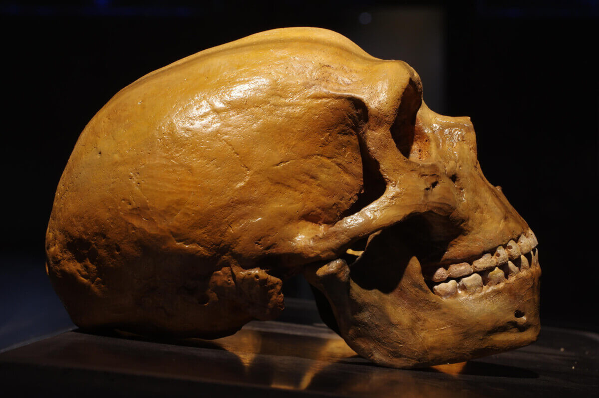
Neanderthal skull vs modern human skull (© Winters860 - stock.adobe.com)
In a Nutshell
- A Neanderthal fetus from Germany had bone structure broadly similar to a late-stage modern human fetus, but the arm and leg bones showed signs of slightly more advanced growth than expected.
- Two baby teeth from other young Neanderthals at the same site contain abnormal gaps in their inner tooth material, representing what the paper describes as one of the earliest, if not the earliest, evidence of metabolic bone disease found in a non-anatomically modern human.
- Fetal bones showed no secondary osteons, a mature bone feature, which supported the estimate that this individual died before or very close to the time of birth, at around eight months of development.
- Researchers stop short of diagnosing any specific condition, and their conclusions lean heavily on comparisons with modern humans, so the true pace of Neanderthal development remains uncertain.
Fifty thousand to ninety thousand years ago, in what is now southeastern Germany, a pregnant Neanderthal lost her baby. Tiny bones left behind, some barely the length of a thumb, sat largely unstudied for decades. A team of researchers has now peered inside those ancient fragments using ultra-precise scanning technology, and what they found is both surprising and deeply human: the bones of this unborn Neanderthal looked broadly similar to those of modern human fetuses, with a few areas that seemed slightly more advanced.
Scientists have long debated just how similar Neanderthals were to modern humans in their earliest days of life. Did their skeletons form the same way? Did their babies develop on the same schedule?
Fossil records of infant and fetal Neanderthals are quite sparse, which makes those questions hard to answer. But a rock shelter in Bavaria called Sesselfelsgrotte, one of the richest Neanderthal sites in western Europe, preserved something rare: the fragmented bones of a Neanderthal who never made it to birth, along with two baby teeth from two other young Neanderthals, including one tentatively identified lower molar.
Those remains, studied with a technique called microcomputed tomography, a high-resolution internal scan that maps the microscopic structure of bone without cutting into it, have offered the clearest window yet into how Neanderthal fetuses developed. Buried inside two of the baby teeth, researchers found what the paper describes as one of the earliest, if not the earliest, cases of metabolic bone disease ever identified in a non-anatomically modern human: evidence of a likely disturbance in mineral metabolism, possibly tied to vitamin D deficiency or another cause such as calcium deficiency, intestinal disease, or kidney problems.
Neanderthal Fetal Bones Under the Microscope
Sesselfelsgrotte 1, as the fetal individual is formally known, left behind 12 bone fragments, some less than 2 centimeters long. Pieces of the skull, jaw, ribs, spine, and limbs make up a partial skeleton that researchers estimated roughly two decades ago belonged to a fetus around eight months along, based on size alone. This new study used scans to examine the bone’s internal architecture: the microscopic channels and tissue patterns that form as a skeleton grows.
What the scans showed matched what appears in a modern human fetus during the final stretch of pregnancy. Bones carried a pattern of rapidly deposited tissue, a scaffold-and-tube structure that floods developing bone with blood supply to fuel quick growth. That same bone formation happens in modern human babies approaching birth, when the skeleton is still racing to build itself. None of the fragments carried the more mature bone patterns that appear after birth, which supported the age estimate.
Leg and arm bones stood out from the skull and jaw bones in a telling way. Long bones, among them the femur, humerus, ulna, and fibula, showed regions of advanced growth compared with the mandible and frontal bone. Researchers note that limb bones and skull bones form through different processes, which could partly explain the gap. Another possibility exists: that Neanderthal limb bones were laying down tissue at a slightly faster rate even in the womb, foreshadowing the rugged, heavily built skeletons Neanderthals are known for as adults.
Researchers are careful to say this cannot be confirmed with the data available. It reads as a possibility, not a conclusion.

Baby Teeth Hint at an Ancient Mineral Metabolism Problem
Two of the other Sesselfelsgrotte individuals, known only from a pair of baby molars, told a different and more troubling story. Inside the hard material that forms the bulk of each tooth, scans revealed clusters of tiny dark spots and gaps, regions where the tooth simply did not mineralize properly. In plain terms, holes sit in the tooth’s inner structure where solid material should be.
Defects like these appear when the microscopic mineral crystals that should fuse together as a tooth forms fail to do so, leaving unmineralized pockets behind. A small amount of this is considered normal. But what researchers found in these Neanderthal teeth sits in the deeper layers of the tooth material, away from where typical developmental defects show up, and clusters in a way consistent with a whole-body disruption at a specific point in time, not a flaw confined to the tooth.
In living patients today, that pattern is associated with disorders of mineral metabolism, including rickets, a condition tied to vitamin D or calcium deficiency that weakens bones and teeth during development. Based on where the defects sit within the teeth, researchers place the disruption somewhere between the last trimester of pregnancy and the middle of the second year of life.
Researchers stop well short of diagnosing these Neanderthals with any specific condition. Causes of this kind of dental defect are many, with nutritional deficiency, intestinal disease, and kidney problems among them, and pinpointing the exact one in fossil remains is not possible. Even so, the paper calls the finding the earliest evidence of metabolic bone disease in a non-anatomically modern human to date, with the tooth from Sesselfelsgrotte 2 coming from a layer dated to around 75,000 years ago by thermoluminescence.

Neanderthal Development Before Birth
Reports of similar tooth defects in Neanderthals have been extremely rare, limited to a tooth from a site in Greece dated to roughly 38,000 to 44,000 years ago, and some disputed cases from a site in Israel. Teeth from Sesselfelsgrotte, if the dating holds, push that evidence back by tens of thousands of years and involve young children rather than adults.
Bone analysis, meanwhile, adds to a growing body of evidence that Neanderthal development in the earliest stages of life closely tracked that of modern humans. A previous study of a young Neanderthal child found that the bone structure in its thigh bone matched that of modern human children several years older, a sign that somewhere between infancy and childhood, Neanderthal bone growth began to diverge and speed up. New findings raise the possibility that the split may have roots reaching back even further, to before birth.
Researchers acknowledge several limits. Scans could not capture the finest level of bone detail, and the team chose not to cut into the bones in order to preserve material for future DNA and dating work. Because the fossils are so fragmentary, only portions of each bone could be examined rather than complete cross-sections. And because the comparisons lean on modern humans as the reference point, the team admits it is hard to say just how different, or how similar, the pace of Neanderthal development truly was.
A portrait emerges from these tiny fragments that is, for all its gaps, vivid. A Neanderthal fetus, eight months along, with a skeleton assembling itself in ways that would look familiar in any modern human developmental study, plus hints of something faster forming in the limbs. Nearby, in two small teeth worn down by a life that ended far too young, a chemical record of hardship set in from the start.
Neanderthals shared the planet with early modern humans, shared genes with them, and, it now seems, shared much of the way their earliest skeletons formed.
Paper Notes
Limitations
Several important constraints frame the study, and the authors are upfront about them. Microcomputed tomography scans, while non-invasive, did not reach the resolution needed to examine bone at the level of individual cells, so certain tissue classifications could not be confirmed with full certainty. Permission was not granted for more detailed invasive analyses, such as cutting the fossils for ground histology, which would have delivered higher-resolution data. Fossils also could not be moved to a more powerful scanning facility, out of concern for radiation exposure and future DNA analysis. Because the bones are highly fragmented, only portions of each skeletal element could be examined rather than complete cross-sections, which limits how much can be said about the full bones. Comparative analysis depends heavily on modern human data, since Neanderthal fetal bone microstructure is almost entirely unstudied, a limitation the authors flag as introducing a degree of circular reasoning when estimating age and identifying differences. Bioerosion in the comparative Neanderthal specimens (La Ferrassie 4bis and Le Moustier 2) also hampered direct comparison. For the teeth, the fragmentary and worn specimens limited the ability to read incremental growth lines and to pinpoint when the mineral defects formed.
Funding and Disclosures
According to the paper, Justyna J. Miszkiewicz receives funding from the Australian Research Council (grant FT240100030). Ricardo Miguel Godinho is funded by the Fundação para a Ciência e a Tecnologia (contract reference 2023.10993.TENURE.006; R&D project ‘ParaFunction’, reference 2022.07737.PTDC). Alvise Barbieri is funded by the Portuguese Ministry of Science (2002.08622.CEECIND) and received funding for the analysis of the Sesselfelsgrotte skeletal and dental remains from the National Geographic Society (NGS-96087R-22). The authors declare no competing interests. They also state that they did not use AI-assisted technologies in creating the article.
Publication Details
Authors: Justyna J. Miszkiewicz, Ricardo Miguel Godinho, Anne Marie Sohler-Snoddy, Kerstin Pasda, Florent Détroit, Patrick Mahoney, Thomas Rathgeber, Cosimo Posth, Thorsten Uthmeier, and Alvise Barbieri.
Journal: Royal Society Open Science, Volume 13, article 260485 (2026).
Paper Title: “Early development of Neanderthals revealed through virtual microanatomy”
DOI: 10.1098/rsos.260485
Received: March 11, 2026. Accepted: May 4, 2026.
Micro-CT scans referenced in the study are available open access via Morphosource at https://www.morphosource.org/organizations/000809898.


