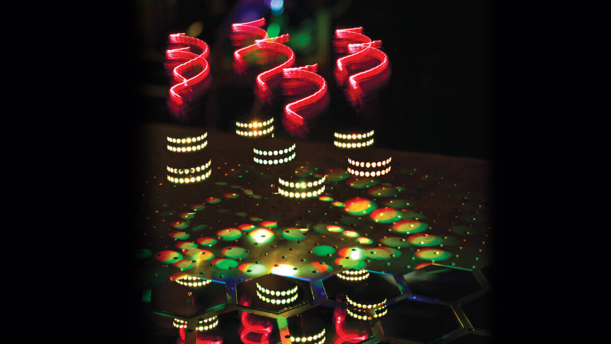
Artistic representation of hyper-Raman optical activity: twisted light (red helices) incident on molecules arranged on a helical scaffold (white dots) produce hyper-Raman scattering spectra (multicoloured light patches) that express ‘chirality’ (patches in spiral patterns and broken mirror). (Credit: Ventsislav Valev and Kylian Valev)
BATH, England — Sometimes, the things we can’t see wind up showing us the world in a way we never imagined. Such is the case after a team of scientific sleuths pulled off the molecular equivalent of catching lightning in a bottle. Led by physicists at the University of Bath, this international band of researchers has coaxed light itself to reveal secrets hidden in the very fabric of matter. Their discovery sounds like something out of a sci-fi novel, but its implications are thrillingly real.
From unmasking counterfeit drugs to sniffing out environmental pollutants, from preserving priceless works of art to pioneering new medical diagnostics, this breakthrough promises to revolutionize how we peer into the microscopic realm. And the kicker? It all hinges on a theory older than many of the scientists who just proved it right.
To understand its significance, we need to start with some basics about how light interacts with molecules.
When light shines on molecules, most of it bounces off unchanged. However, for every million light particles (photons), one changes color. This change, known as the Raman effect, helps scientists identify molecules and understand their energy states. But some molecular features remain invisible to this technique.
Enter hyper-Raman scattering. This more advanced phenomenon occurs when two photons simultaneously hit a molecule and combine to create a single scattered photon with a color change. Hyper-Raman has several advantages over regular Raman scattering: it can penetrate deeper into living tissue, is less likely to damage molecules, and produces clearer images with less background noise.
Despite these benefits, hyper-Raman has been unable to study a crucial property of many biological molecules: chirality. Chirality refers to a molecule’s “handedness” or sense of twist, similar to the helical structure of DNA. Many important biomolecules, including proteins, RNA, sugars, and some vitamins, exhibit chirality.
The 45-year-old theory that shed ‘light’ on the discovery
In 1979, researchers David L. Andrews and Thiruiappah Thirunamachandran theorized that using chiral light for hyper-Raman scattering could reveal three-dimensional information about molecules, including their chirality. However, this effect, dubbed “hyper-Raman optical activity,” was expected to be so subtle that many scientists doubted it could ever be measured.
The Bath-led team took an innovative approach to overcome this challenge. Instead of trying to measure the effect directly from chiral molecules, they used an indirect method. They deposited non-chiral molecules onto tiny gold nanohelices – spiral-shaped structures about 100 times smaller than the width of a human hair. These nanohelices effectively transferred their twist (chirality) to the molecules.
“While previous attempts aimed to measure the effect directly from chiral molecules, we took an indirect approach,” explains University of Bath Professor Ventsislav Valev, who led the study, in s statement. “We employed molecules that are not chiral by themselves, but we made them chiral by assembling them on a chiral scaffold. Specifically, we deposited molecules on tiny gold nanohelices that effectively conferred their twist (chirality) to the molecules.”
This ingenious approach had an additional benefit. The gold nanohelices acted as tiny antennas, focusing light onto the molecules and amplifying the hyper-Raman signal, making it detectable.
What hyper-Raman optical activity means for the future
The successful demonstration of hyper-Raman optical activity opens up exciting new possibilities for molecular analysis. It could help pharmaceutical companies analyze and control the quality of drugs, assist in identifying counterfeit products, detect illegal drugs and explosives at crime scenes or customs checkpoints, and reveal pollutants in environmental samples.
In the art world, this technique could aid in the conservation and restoration of paintings by revealing the composition of pigments. Medical researchers might use it to detect disease-induced molecular changes for diagnostic purposes.
Emeritus Professor Andrews from the University of East Anglia, one of the original theorists who predicted this effect, expressed his satisfaction with the results: “It is very gratifying to see this work the experimental finally confirm our theoretical prediction, after all these years. The team from Bath have performed an outstanding experiment.”
While this breakthrough marks a significant milestone, there’s still a long road ahead before hyper-Raman optical activity becomes a standard analytical tool. The researchers are looking forward to this journey, collaborating with Renishaw PLC, a world-renowned manufacturer of Raman spectrometers, to further develop and refine the technique.
Discoveries like this remind us of the vast potential still waiting to be unlocked in the world of molecular science. By providing a new way to “see” molecules, this research opens up exciting avenues for scientific exploration and technological innovation across a wide range of fields.
“This research work has been a collaboration between chemical theory and experimental physics across many decades and across academics of all stages – from PhD student to Emeritus Professor,” says Valev. “We hope it will inspire other scientists and that it will raise awareness that scientific progress often takes many decades.”
Paper Summary
Methodology
The researchers created arrays of gold nanohelices using a technique called nano glancing angle deposition. They then coated these spirals with crystal violet, a common dye molecule. The team used a specially modified Raman spectrometer that could produce and detect circularly polarized light at both the fundamental frequency (1064 nm) and the second harmonic (532 nm). They illuminated the samples with left- and right-handed circularly polarized light and measured the differences in the scattered light to detect the hyper-Raman optical activity effect.
Results
The key finding was that the hyper-Raman spectra of the crystal violet molecules showed different intensities when illuminated with left- versus right-handed circularly polarized light. Importantly, this difference changed sign when the handedness of the underlying gold nanohelices was switched from left to right. This demonstrates that the chirality of the nanohelices was indeed transferred to the normally achiral dye molecules, enabling the observation of hyper-Raman optical activity.
Limitations
One challenge in this study was the subtlety of the hyper-Raman optical activity effect, which had made it difficult to observe in previous attempts. The researchers overcame this by using the gold nanohelices to both confer chirality and amplify the signal. However, this indirect approach means that further work may be needed to observe the effect directly in naturally chiral molecules. Additionally, the current setup is complex and will require further development before it can become a widely accessible analytical tool.
Discussion and Takeaways
This study demonstrates a new way to probe molecular structures and properties using light. By confirming a decades-old theoretical prediction, it opens up new possibilities for studying chirality in molecules, which is crucial in many biological and chemical processes. The technique could have wide-ranging applications, from pharmaceutical quality control to environmental monitoring and medical diagnostics. However, the researchers emphasize that this is just the first step, and significant work remains to develop this into a standardized analytical method.
Funding and Disclosures
The research was supported by various grants from organizations including The Royal Society, the Leverhulme Trust, and the Engineering and Physical Sciences Research Council (EPSRC). The researchers also noted their collaboration with Renishaw PLC, a manufacturer of Raman spectrometers, in developing this technique further.


