
Credit: PeopleImages on Shutterstock
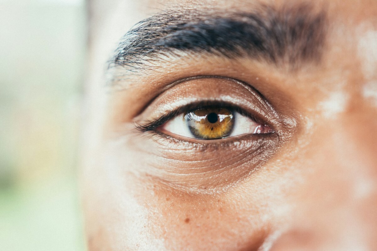
Scientists suggest a simple eye scan could potentially help prevent cognitive decline
In A Nutshell
- Researchers achieved 94% accuracy in identifying Alzheimer’s disease by analyzing bacterial infections, inflammatory markers, and toxic proteins in donated eye tissue, though this has not yet been validated in living patients.
- Eyes of Alzheimer’s patients contained about three times more Chlamydia pneumoniae bacteria than those with normal cognition, and the infection appears to trigger inflammation that worsens brain pathology.
- Machine learning models could predict disease severity based purely on eye findings, raising the possibility of future non-invasive screening tests that catch Alzheimer’s before symptoms appear.
- Some cognitively healthy people showed high bacterial and inflammatory markers in their eyes, suggesting a potential window for early intervention, though more research is needed to determine if this predicts future decline.
Doctors are exploring whether a simple eye exam could one day predict Alzheimer’s disease, no brain scan or spinal tap required.
Researchers at Cedars-Sinai Medical Center have developed a method that combines eye imaging with artificial intelligence to spot warning signs of dementia in donated tissue samples, achieving 94% accuracy in distinguishing between people who had normal cognition, mild cognitive impairment, or Alzheimer’s at death. That accuracy comes from analysis of postmortem eye tissue using a small test set of 14 subjects and has not yet been validated in living patients.
The finding offers a potential path toward transforming how we catch Alzheimer’s. Right now, by the time someone notices memory problems, their brain has already suffered significant damage. If researchers can develop ways to detect these same markers in living eyes, it could identify at-risk people while there’s still time to intervene. For instance, with medications that slow or prevent disease progression.
A Surprising Culprit Hiding in Plain Sight
The study of 104 deceased donors uncovered the eyes of Alzheimer’s patients harbored about three times more of a common respiratory bacteria in their retinas (and four times more in matched brain tissue) compared to people with normal cognition. The culprit: Chlamydia pneumoniae, the same bug that causes pneumonia and sinus infections.
Dr. Maya Koronyo-Hamaoui’s team found bacterial clusters nestled inside retinal cells, particularly in people with dementia. Even more intriguing, some cognitively healthy people showed high bacterial levels in their eyes, suggesting the infection can be present before symptoms appear, though not everyone with these signs goes on to develop dementia.
Next, when researchers infected human brain cells with C. pneumoniae in the lab, the cells produced several-fold more of the sticky amyloid-beta protein that forms plaques in Alzheimer’s brains. Infected cells also showed signs of severe damage, dying at rates nearly four times higher than uninfected cells.
In mice already genetically prone to Alzheimer’s-like disease, a single bacterial infection made everything worse. Six months after exposure, infected mice had more brain plaques and performed poorly on memory tests compared to uninfected mice with the same genetic risk.
The bacteria doesn’t just sit there: it actively fans the flames of disease by triggering an inflammatory alarm system in cells. This creates a vicious cycle. Inflammation damages tissue, which attracts more immune cells, which causes more inflammation. Meanwhile, the brain’s cleanup crew (cells called microglia that normally clear out debris and pathogens) gets overwhelmed and can’t keep up.
How the Test Works
The breakthrough came from machine learning. Researchers trained AI models to analyze eye tissue for multiple factors: bacterial load, inflammatory markers, and toxic proteins. Looking at bacterial levels alone gave moderate results. But combining those numbers with measurements of amyloid-beta in the retina pushed accuracy to 94% for distinguishing between the three groups in their dataset.
Performance dropped when identifying mild cognitive impairment specifically, highlighting the need for larger, longitudinal studies. The models could also predict disease severity based purely on eye findings. Higher bacterial loads correlated with more brain shrinkage and worse cognitive test scores.
From Lab to Clinic
Whether these findings, published in Nature Communications, can translate into routine eye exams for living patients remains an open question. Researchers need to develop technology that can detect these markers non-invasively. Several research groups are working on specialized retinal cameras and fluorescent dyes that light up problem proteins. Some have successfully imaged amyloid deposits in living human eyes.
The timing question looms large. Three women in the study had normal cognition but showed the bacterial and inflammatory patterns typically seen in Alzheimer’s patients. Their brains also had elevated disease-related pathology despite their intact memory and thinking skills. This raises the possibility that eye scans might catch people on the cusp of decline (a critical window for treatment) though more research in living patients is needed to confirm this.
Treatment options may already exist. A large observational study in Taiwan found that pneumonia patients who received appropriate antibiotics for C. pneumoniae infections had lower rates of Alzheimer’s later in life compared to those who weren’t adequately treated. The study does not prove that antibiotics prevent Alzheimer’s, only that treated infections were linked to lower observed risk. Azithromycin, commonly prescribed for respiratory infections, showed particularly promising associations.
Drug companies are also developing medications that block the inflammatory alarm system triggered by the bacteria. Several of these anti-inflammatory drugs are already in clinical trials for other diseases.
Eye Scans Could Flag Bacteria, Prevent Dementia
About 80% of people over age 60 carry antibodies against C. pneumoniae, meaning they’ve been exposed at some point. Yet not everyone develops dementia. The bacteria likely acts more like an accelerant than a match: it amplifies damage in people already vulnerable due to age, genetics, or early protein buildup in their brains.
This means blanket antibiotic treatment doesn’t make sense and could fuel antibiotic resistance. Instead, doctors need better ways to identify which patients have active infections driving their disease and who would actually benefit from treatment.
That’s where eye imaging could help, if the technology can be validated in living patients. Researchers are now planning studies that follow people over time, tracking how eye changes predict who develops dementia and who stays healthy.
If those studies pan out, eye scans could one day complement other screening tools, serving as a simple addition to routine care that might catch serious trouble early, when there’s still time to do something about it.
Disclaimer: This article reports on early-stage research conducted using postmortem tissue samples. The findings have not yet been validated in living patients, and no clinical diagnostic test based on this research is currently available. The study’s machine learning models were developed and tested on small sample sizes. Readers should not interpret these findings as immediately actionable medical advice. Anyone concerned about Alzheimer’s risk should consult with their healthcare provider about currently available screening and prevention strategies.
Paper Notes
Study Limitations
Sample sizes for some analyses remained relatively small, particularly for brain tissue specimens and the mild cognitive impairment group. All human data came from postmortem examinations, preventing researchers from tracking changes over time or establishing definitive causality. Machine learning models showed reduced accuracy for identifying mild cognitive impairment compared to Alzheimer’s dementia or normal cognition. Researchers could not determine when infections initially occurred or whether bacterial loads reflected recent versus decades-old exposures. The machine learning test set was small with only 14 subjects, limiting the generalizability of predictive accuracy findings.
Funding and Disclosures
Research support came from the National Institutes of Health/National Institute on Aging grants R01AG056478, R01AG055865, AG056478-04S1, and R01AG075998, plus Alzheimer’s Association grant AARG-NTF-21-846586. Additional funding came from the Goldrich and Snyder Foundations and the Ray Charles Foundation. Co-authors Yosef Koronyo, Keith L. Black, and Maya Koronyo-Hamaoui are co-founders of NeuroVision Imaging, Inc., which develops retinal imaging technologies. NeuroVision Imaging had no role in study design, data collection, analysis, or manuscript preparation.
Publication Details
Authors: Bhakta Prasad Gaire, Yosef Koronyo, Jean-Philippe Vit, Alexandre Hutton, Lalita Subedi, Dieu-Trang Fuchs, Natalie Swerdlow, Altan Rentsendorj, Saba Shahin, Daisy Martinon, Edward Robinson, Alexander V. Ljubimov, Julie A. Schneider, Lon S. Schneider, Debra Hawes, Stuart L. Graham, Vivek K. Gupta, Mehdi Mirzaei, Keith L. Black, Jesse G. Meyer, Moshe Arditi, Timothy R. Crother, and Maya Koronyo-Hamaoui | Journal: Nature Communications | Publication Date: January 22, 2026 | Volume: 17, Article Number: 771 | DOI: https://doi.org/10.1038/s41467-026-68580-4 | Institutions: Cedars-Sinai Medical Center (Los Angeles, CA), Macquarie University (Sydney, Australia), Rush University Medical Center (Chicago, IL), University of Southern California (Los Angeles, CA), and University of California Los Angeles


