(credit: Sherbak_photo/Shutterstock)
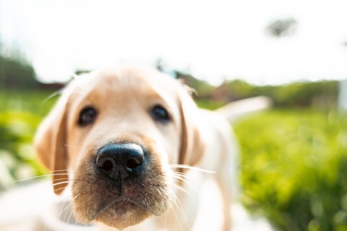
GENEVA, Switzerland — Have you ever wondered why dogs, cows, and other mammals have those distinctive geometric patterns on their wet noses? Those polygonal shapes aren’t just random decorations – they’re intricate structures that help keep animals’ noses moist and aid in their ability to smell and regulate temperature. Now, fascinating research shows us the remarkable process behind how these patterns form during embryonic development, challenging our understanding of how complex biological structures emerge.
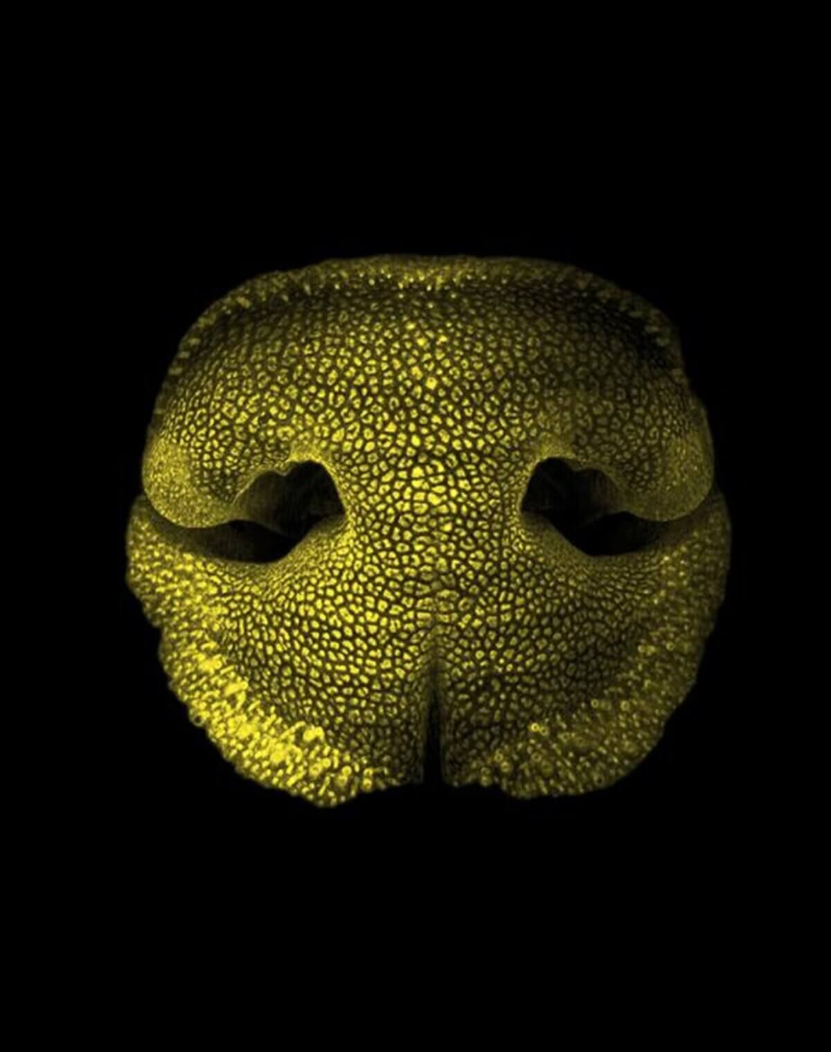
The study, led by researchers at the University of Geneva and several other institutions across Europe, discovered that these nose patterns – technically called “rhinoglyphics” – form through a fascinating mechanical process rather than being directly encoded in DNA. It’s similar to how cracks form in drying mud, but with an ingenious biological twist that ensures the patterns develop precisely where they’re needed.
This finding, published in Current Biology, is particularly exciting because it reveals a new principle in developmental biology: the concept of “mechanical positional information.” Just as GPS coordinates tell you where you are on Earth, developing tissues need information about where to form specific structures. Scientists have long known that chemical signals serve this purpose, but this study shows that mechanical forces can also provide this crucial positional information.
“Finding specific examples of beautiful patterns in living organisms is easy,” explains study co-author Michel Milinkovitch, a professor in Geneva’s Department of Genetics and Evolution, in a statement. “All we have to do is look around us! Our latest study focuses on the noses of dogs, ferrets and cows, which exhibit a singular network of polygonal structures.”
The Nose Knows: Form Follows Function
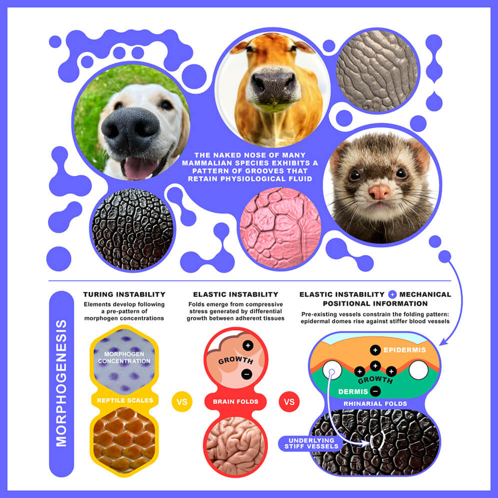
These polygonal patterns aren’t just for show. In dogs, they help collect and retain moisture that keeps their noses wet, which is crucial for their legendary sense of smell. The moisture helps trap scent molecules, which can then travel to special sensory organs. In cows, these patterns include tiny ducts that secrete fluid directly onto the nose’s surface. The patterns also play a role in temperature regulation – in rodents, they help cool the brain, while in carnivores, a cool nose might help them detect warm prey through infrared sensing.
To understand how these patterns form, the research team examined nose development in cow, dog, and ferret embryos using advanced imaging techniques. They discovered that the process happens in three main stages, like a carefully choreographed architectural project.
The Blueprint: How Nature Builds a Patterned Nose
First, a network of blood vessels forms beneath the skin. These vessels serve as a template, much like the foundation of a building. Next, the base layer of the skin (called the epidermis) begins to fold, forming cup-like structures between the blood vessels. Finally, the outer skin layer develops creases that align perfectly with the underlying blood vessels, creating the distinctive polygonal domes we see on adult animals’ noses.
What makes this process remarkable is that it’s self-organizing – meaning these patterns emerge naturally from the physical properties of the growing tissues, rather than following a strict genetic blueprint. It’s similar to how soap bubbles naturally form hexagonal patterns when squeezed together, but with an important difference: the blood vessels act as guide posts, ensuring the patterns form in exactly the right places.
The researchers confirmed this mechanism through both detailed observations and computer simulations. They created virtual models of growing nose tissue and showed that when the blood vessels were made stiffer than the surrounding tissue – as they are in real animals – the patterns formed correctly. When this stiffness difference was removed in the simulation, the patterns became irregular and unrealistic.
“Our numerical simulations show that the mechanical stress generated by excessive epidermal growth is concentrated at the positions of the underlying vessels, which form rigid support points,” explains first author Paule Dagenais, post-doctoral fellow in the Department of Genetics and Evolution. “The epidermal layers are then pushed outwards, forming domes – akin to arches rising against stiff pillars.”
A Natural Experiment: Clone Comparison
One of the most compelling pieces of evidence came from comparing the nose patterns of cloned cows. Despite having identical DNA, these clones showed as much variation in their nose patterns as unrelated cows. This proves that the exact arrangement of the patterns isn’t genetically predetermined but emerges through this mechanical process.
The research team also examined cows with a condition called Ehlers-Danlos syndrome, which affects tissue elasticity. These cows showed disrupted nose patterns, further confirming that mechanical properties, rather than genetic instructions alone, are crucial for proper pattern formation.
Understanding how mammal noses develop their distinctive patterns does more than satisfy scientific curiosity. The research reveals new principles of biological development that could inspire innovations in tissue engineering and biomaterial design, showing once again how nature’s solutions can guide human innovation.
“This is the first time that mechanical positional information has been described to explain the formation of structures during embryonic development,” concludes Milinkovitch. “But we are confident that it will help explain the formation of other biological structures associated to the presence of blood vessels.”
Paper Summary
Methodology
The researchers used multiple sophisticated techniques to study nose development. They collected embryos from cows, dogs, and ferrets at different developmental stages and used special microscopy methods that allowed them to see through the tissue in three dimensions. They combined this with traditional tissue staining techniques to identify different cell types and track cell division. Computer simulations were used to test their hypotheses about how mechanical forces shape the developing patterns. They also used 3D scanning techniques to compare nose patterns between different animals, including cloned cows.
Key Results
The study found that nose patterns form through a three-stage process involving blood vessels, skin folding, and surface crease formation. The patterns emerge through mechanical self-organization but are guided by the stiffness of blood vessels. Cell proliferation was found to be uniform across the tissue, ruling out the possibility that localized cell growth creates the patterns. Cloned cows showed similar levels of pattern variation as unrelated cows, demonstrating that exact pattern positions aren’t genetically determined.
Study Limitations
The researchers note that their computer models used simplified mechanical properties and couldn’t account for all biological complexities. They couldn’t directly measure the stiffness of embryonic tissues, instead using cell density as an approximate indicator. The study focused on three species, so the findings might not apply to all mammals with similar nose patterns.
Discussion & Takeaways
This research reveals a new principle in developmental biology where mechanical properties of tissues can provide positional information for pattern formation. This is different from the traditional view where chemical signals guide development. The findings might help explain other biological patterns and could have applications in tissue engineering and biomaterial design. The study also suggests that similar mechanical principles might be involved in other biological patterns, such as fingerprints.
Funding & Disclosures
The research was supported by multiple organizations including the Swiss National Science Foundation, the European Research Council, and the International Human Frontier Science Program Organization. The authors declared no competing interests. The study complied with all relevant ethical guidelines and regulations for animal research.


