
(© Gorodenkoff - stock.adobe.com)
In a Nutshell
- Researchers mapped more than 3 million cells from 527 women aged 15 to 86 and found that breast tissue undergoes a broad, sweeping decline with age, losing cells, structural density, and cellular activity across virtually every cell type.
- The immune environment of breast tissue shifts with age from one rich in antibody-producing B cells to one dominated by inflammatory and tissue-repair cells, a change that may help explain why breast cancers in younger and older women behave so differently.
- The physical architecture of breast tissue is dramatically overhauled with age, with far fewer milk-producing lobules, more and larger fat cells, reduced blood vessels, and a weakening of the spatial organization that holds healthy tissue together.
- Menopause emerged as the single dominant driver of breast tissue aging, producing one massive wave of change in a woman’s late 40s rather than the gradual, multi-stage process seen in other parts of the body.
Conventional thought holds that aging is a gradual, not always gentle process. But a sweeping new study of breast tissue tells a different story: as women get older, their breast tissue doesn’t just slowly shift as other parts of the body do. It loses cells, loses structure, and reshapes itself so thoroughly that researchers describe it as “an unexpected general decline.” The transformation may help explain why breast cancer behaves so differently in younger women compared to older ones.
An international team of researchers analyzed more than 3 million cells from 527 women who had undergone breast reduction surgeries. The women ranged in age from 15 to 86, with a median age of 38, making this one of the largest and most detailed surveys of normal breast tissue ever conducted. Using a technology that can measure 40 different proteins at once at a level finer than a single cell, the team built a first-of-its-kind atlas of how the breast transforms across a lifetime.
A Breast Tissue Atlas Built on 3 Million Cells
Working out of the University of Cambridge and the BC Cancer Agency in Vancouver, the research team used a technique that fires a tiny laser at tissue samples and employs a specialized detector to measure dozens of proteins in each cell simultaneously. The result is a detailed portrait of what kinds of cells are present, what they are doing, and how they are arranged relative to one another.
Tissue samples from breast reduction surgeries unrelated to cancer were organized on small glass slides, each containing tissue cores from individual patients. The team imaged a total of 1,710 cores, and a breast pathologist reviewed the images to label structural features. From the data, they identified 25 distinct cell types, including 11 types of the cells that line the breast’s ducts and milk-producing units, along with 14 types of surrounding cells such as immune cells, blood vessel cells, and connective tissue cells. They then tracked how the abundance, behavior, and spatial arrangement of all these cells changed across the full age range.
One of the most dramatic findings of the study, published in Nature Aging, was how universally breast tissue slows down with age. Cell density, or how tightly packed the tissue is, declined for nearly every category of cell. And the decline was not limited to just having fewer cells. Across almost every single cell type, the fraction of cells actively dividing also dropped with age.
The cells lining ducts and milk-producing units showed the steepest drop in division rates, but immune and connective tissue cells followed close behind. Even cell size changed: connective tissue cells steadily shrank as women aged. Dividing cells tend to be larger than non-dividing ones, which makes sense because a cell roughly doubles in size before splitting in two. But even after accounting for that, the shrinkage held.
How Aging Reshapes Breast Tissue Architecture
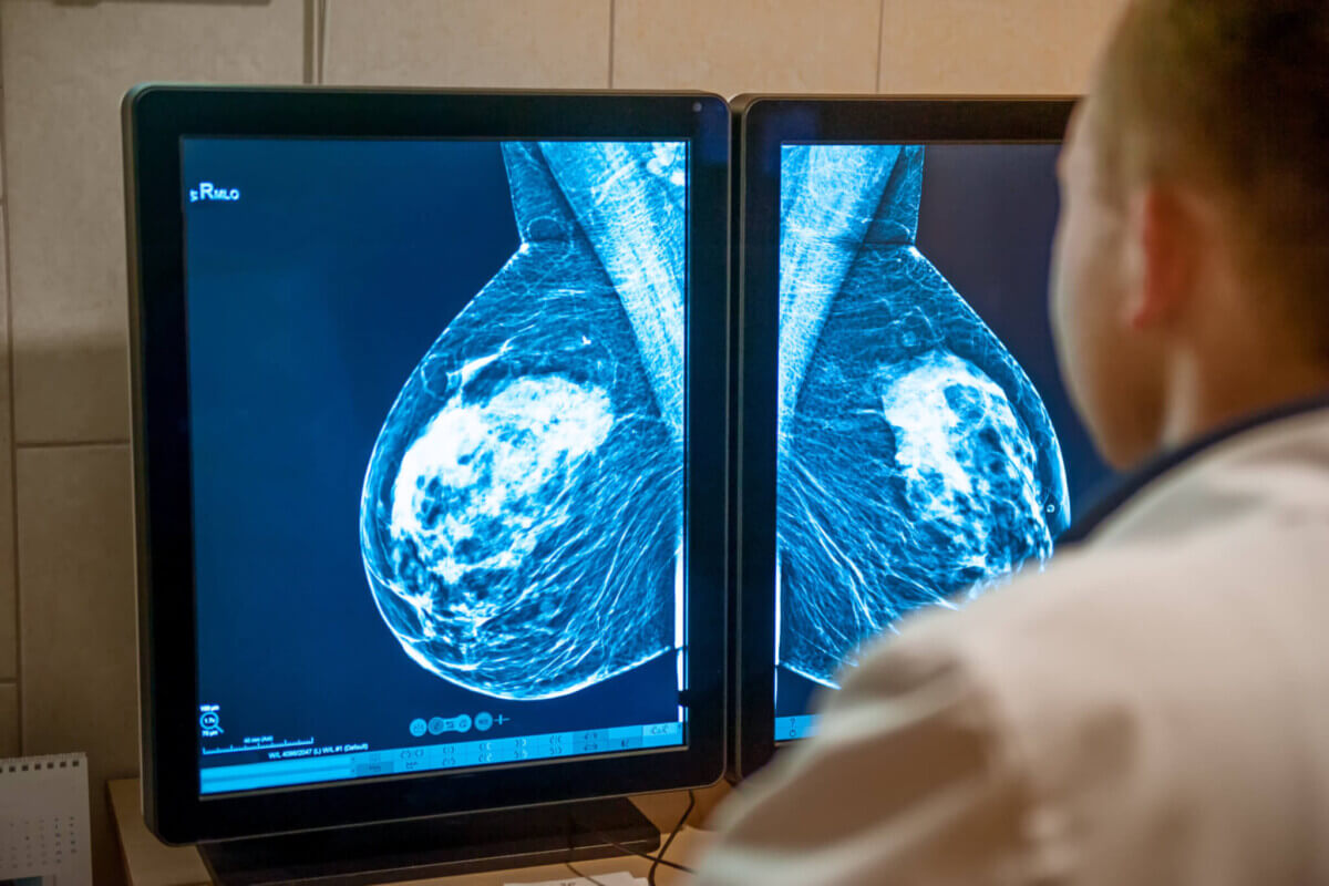
Beyond individual cells, the physical architecture of breast tissue was overhauled. Lobules, the small grape-like clusters of cells responsible for milk production, plummeted with age, with a particularly sharp drop around age 50, roughly corresponding to menopause. Meanwhile, the proportion of tissue occupied by fat cells grew, and fat cells themselves got bigger. Blood and lymphatic vessels took up less space in older tissue as well.
One unexpected twist: the specialized layer of cells that wraps around ducts and lobules, a barrier thought to help contain early-stage cancers, actually got thicker with age in both structures. That ran counter to the overall pattern of decline.
The researchers also looked at how different cell types were physically positioned relative to one another. In older tissue, several cell types, including blood vessel cells, a type of immune cell involved in tissue repair, and connective tissue cells, were all located farther from the cells lining ducts and lobules. No cell type moved closer with age. The structural scaffolding that keeps healthy tissue organized appeared to weaken over time.
The immune system’s presence in breast tissue shifted too. B cells, which produce antibodies, were by far the most common immune cell in younger tissue. In older tissue, a specific type of killer T cell armed with a cell-destroying protein was more common, along with immune cells associated with inflammation and tissue repair rather than direct germ fighting. When the researchers grouped cells into larger multicellular neighborhoods, clusters of cells that tend to appear together, they found ten recurring patterns. Most of these neighborhoods were less dense in older tissue. The most dramatic depletion was in neighborhoods rich in lobules and actively dividing cells, reinforcing the picture of tissue steadily winding down.

Menopause Emerges as the Single Biggest Driver of Breast Tissue Aging
Using a statistical approach, the team searched for periods during adult life when breast tissue changes most rapidly. Breast tissue appeared to be dominated by a single massive wave of change in the late 40s, corresponding to menopause. A smaller peak appeared in the 20s, driven partly by shifts in certain immune cell populations, but it was dwarfed by the menopausal wave. Hormone-sensitive organs like the breast may not follow the same aging pattern seen elsewhere in the body.
That distinction matters for understanding breast cancer. Tumors do not develop in isolation. They emerge within a specific tissue environment that can either help or hinder their growth. The fact that a 30-year-old’s breast tissue looks fundamentally different from a 60-year-old’s, not just in cell types but in spatial arrangement, immune makeup, and structural organization, could help explain why breast cancers in younger women tend to behave differently from those in older women.
The researchers also explored the relationship between the hormone receptor for estrogen and cell division. They found that most dividing cells lining ducts and lobules were negative for the estrogen receptor. But individual cells that were positive for the receptor and located in lobules were actually more likely to be dividing, a wrinkle that contradicts some earlier research on the topic.
This atlas does not answer every question about why breast cancer risk changes with age. But by mapping normal breast tissue across decades of life at extraordinary resolution, the study reveals that the tissue a tumor is born into is not a fixed backdrop. It is a living, shifting environment that transforms as women age, with the sharpest transformation happening right around menopause.
Disclaimer: This study was conducted using tissue from breast reduction surgeries, which may not fully represent all women. Tissue samples were taken as small cores, which could limit how completely the broader architecture was captured. Cell types were identified based on protein patterns, shape, and location rather than direct functional testing, so the labels may not reflect the full behavior of each cell type. These findings describe associations between aging and breast tissue changes and do not establish cause and effect.
Paper Notes
Limitations
The study relied on tissue from breast reduction surgeries and other noncancer-related procedures, which may not perfectly represent the general population. The tissue was sampled using small cores from each patient, potentially limiting how well the full architecture was captured. The researchers ran additional analyses and found that images greater than 600 micrometers reliably represented tissue features and that their dataset was well powered for evaluating relationships between tissue structure and age. However, they acknowledged that methods for defining cell boundaries face built-in challenges without a strong marker of the cell’s outer edge, and that their measurements probably underestimate cell size. Cell type labels were based on protein profiles, shape, and spatial location but were not confirmed using functional tests, meaning the labels may not entirely capture each cell type’s behavior. The study also noted that variation was greater among the cells lining ducts and lobules than among surrounding cell types.
Funding and Disclosures
Funding for this research came from multiple sources. Senior author H. Raza Ali was supported by a Cancer Research UK core award and an ERC Starting Grant funded by UKRI. Co-first author Pulkit Gupta was supported by the Vanderbilt Medical Scientist Training Program. Senior author Samuel Aparicio holds the Nan and Lorraine Robertson Chair in Breast Cancer and a Canada Research Chair in Molecular Oncology. Additional support came from the Terry Fox Research Institute, the Canadian Institutes of Health Research, the Breast Cancer Research Foundation, the Canada Foundation for Innovation, and BC Cancer Foundation core funding. Co-first author Eric Lee was supported by a CIHR CGS-Doctoral scholarship. The authors declared no competing interests.
Publication Details
Title: Single-cell spatial atlas of the aging human breast
Authors: Pulkit Gupta, Eric Lee, Neus Masqué Soler, Ellen Schrader, Xiao Qian Wang, Shimrit Mayer, Cristina Flores, Sean Beatty, Andrew Roth, Samuel Aparicio, and H. Raza Ali. Pulkit Gupta and Eric Lee contributed equally as co-first authors; Samuel Aparicio and H. Raza Ali jointly supervised the work.
Institutions: CRUK Cambridge Institute, University of Cambridge, Cambridge, UK; Department of Molecular Oncology, BC Cancer Agency, Vancouver, British Columbia, Canada; Department of Pathology and Laboratory Medicine, University of British Columbia; Department of Computer Science, University of British Columbia; Department of Medical Genetics, University of British Columbia; Department of Histopathology, Addenbrookes Hospital, Cambridge, UK.
Journal: Nature Aging
DOI: https://doi.org/10.1038/s43587-026-01104-3
Received: July 24, 2025; Accepted: March 2, 2026


