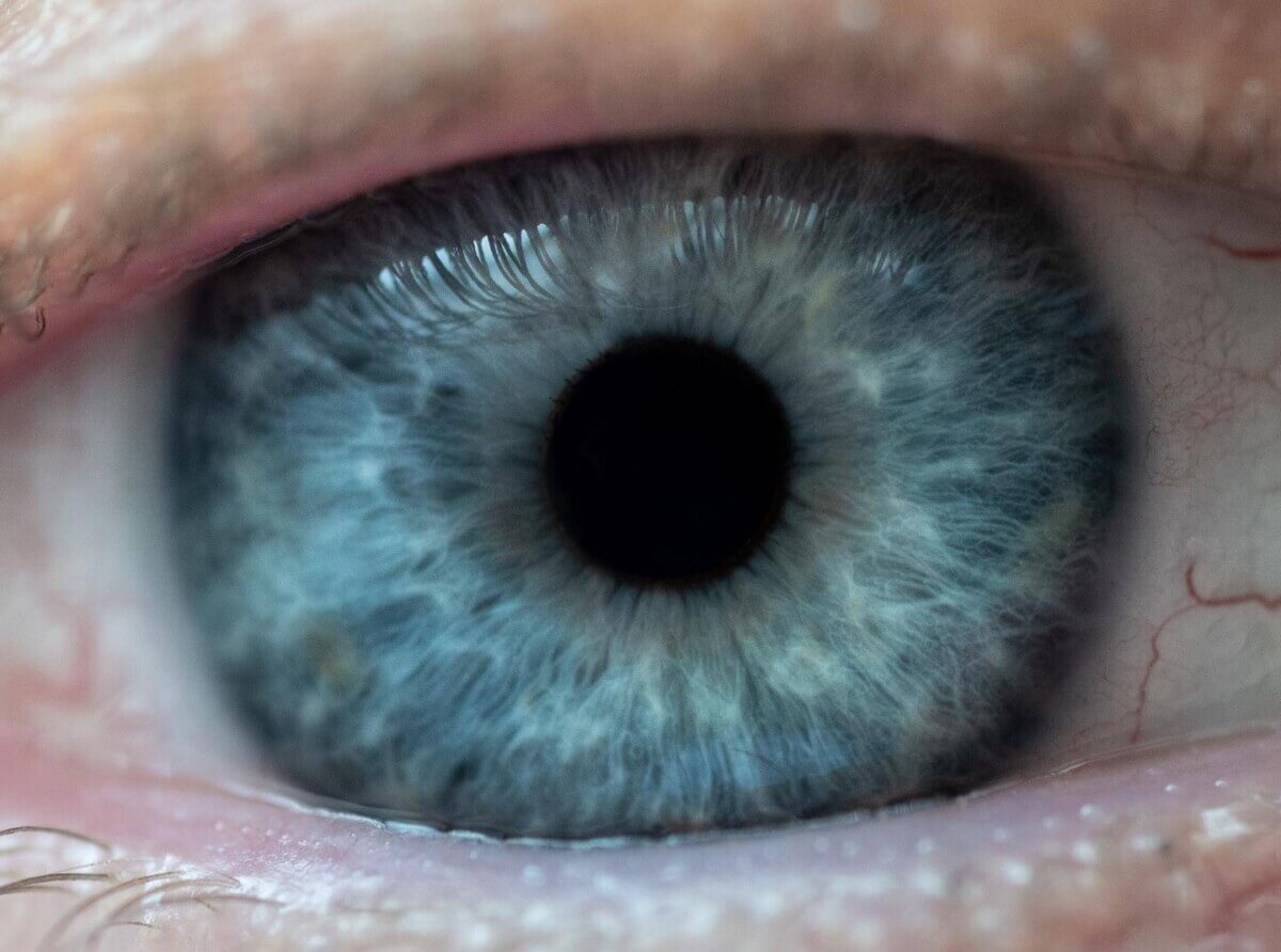
(Credit: Rob Felt, Georgia Tech)
In a Nutshell
- In two mouse models of glaucoma, the cellular system responsible for clearing damaged mitochondria broke down before nerve cells in the eye began dying, suggesting this breakdown is a cause rather than a side effect of the disease.
- When scientists genetically disabled the cleanup system specifically in the eye’s nerve cells, those cells accumulated damaged mitochondria and died, mimicking glaucoma even without elevated eye pressure.
- A drug called Torin 2, which restarts the cellular cleanup process, protected nerve cells and preserved measures of function in mouse models of glaucoma, and improved nerve cell survival in human retinal tissue tested in the lab.
Glaucoma is supposed to be manageable. Lower the pressure inside the eye, protect the vision, and that has been the standard playbook for decades. But for some people living with the disease, vision loss continues even after doctors successfully bring that pressure down. Something else is destroying the eye’s nerve cells, and researchers have been working to understand what it is. New evidence points to a culprit hiding inside the cells themselves: broken cellular cleanup machinery that lets damaged power generators pile up until they become toxic.
Nerve cells that carry visual signals from the eye to the brain are extraordinarily energy-hungry. They rely heavily on mitochondria, the tiny structures that produce energy inside cells, to keep firing. When those mitochondria get damaged, healthy cells have a built-in system to break them down and clear them out, much like a recycling program. A study published in Molecular Neurodegeneration found that in glaucoma, that recycling system breaks down early in the disease, long before the nerve cells themselves start dying. Damaged mitochondria pile up, generate toxic stress, and eventually push the nerve cells past the point of no return.
When the researchers used a drug called Torin 2 to restart the recycling process in mouse models of glaucoma, it prevented nerve cell death and preserved measures of visual function. That same drug also improved nerve cell survival in samples of human retinal tissue kept alive in the lab.
What’s Going Wrong Inside Glaucoma-Damaged Eyes
Glaucoma affects nearly 80 million people worldwide, with that number projected to reach 111.8 million by 2040. It causes irreversible blindness by progressively destroying the nerve cells at the back of the eye and the fibers connecting the eye to the brain. Elevated pressure inside the eye, caused by a drainage problem in the eye’s filtration tissue, is the main known risk factor. But nerve cell damage continues in patients whose eye pressure is well controlled, frustrating researchers and patients alike for years.
This study examined whether a failure of cellular recycling could explain that gap. These particular nerve cells are especially vulnerable because they are long-lived, cannot be replaced, and burn through enormous amounts of energy. All that energy consumption produces a lot of cellular waste and damaged mitochondria, which makes a working cleanup system essential.
Researchers used two mouse models designed to mimic human glaucoma. In one, mice received weekly steroid injections around the eye to raise pressure over 10 weeks, closely resembling a form of glaucoma seen in humans who use steroid medications. In the second, a genetic manipulation triggered the production of a defective protein in the eye’s drainage tissue, causing sustained pressure elevation and mimicking a hereditary form of the disease.
Across both models, chronically elevated eye pressure led to a buildup of mitochondria inside the degenerating nerve fibers. Under a powerful microscope, those mitochondria looked swollen and internally disorganized, consistent with damage and dysfunction. Chemical markers of DNA damage were also elevated in the nerve cell layer.
To measure the recycling system’s activity directly, the team used a specially engineered line of mice with a sensor that glows different colors depending on whether mitochondria are being actively broken down or just sitting idle. Recycling activity dropped after just five weeks of elevated pressure, a point at which nerve cell loss had not yet begun. At 10 weeks, when nerve cells were actively dying, the recycling system was still suppressed. That timing strongly suggested the cleanup failure was happening first, setting the stage for nerve cell death rather than resulting from it.
Proof the Cleanup Failure Drives the Damage
To confirm that the recycling failure caused nerve cell death rather than simply accompanying it, the team used a gene-editing approach to disable the cleanup machinery exclusively inside the eye’s nerve cells, without altering eye pressure. Mice with this targeted change accumulated damaged mitochondria, showed signs of oxidative DNA damage, and lost roughly 39% of their nerve cells along with approximately 59% of healthy optic nerve fibers within six weeks. Disabling the cleanup system alone was enough to reproduce the core features of glaucoma’s nerve damage.
A Drug That Restarts the Cellular Cleanup System
With the mechanism established, the team tested whether pharmacologically restarting the cleanup process could protect nerve cells. Torin 2 works by releasing a molecular brake that normally keeps the recycling system switched off. A single injection into the eye boosted recycling activity within 24 hours in mice. Researchers then gave the drug as a treatment to animals that had already been under elevated pressure for seven weeks.
In the steroid-induced glaucoma model, animals that received Torin 2 scored substantially better on a test of nerve cell function than control animals. Nerve cell counts and healthy fiber counts were also substantially higher in treated eyes, and microscopy confirmed that damaged mitochondria had accumulated far less in those eyes. Treatment also better preserved the process by which material moves along nerve fibers toward the brain.
Similar protective effects appeared in the second mouse model, though the authors noted that in that group, Torin 2 also appeared to reduce eye pressure by promoting the breakdown of the defective protein clogging the drainage tissue. That made it harder to fully separate the drug’s direct nerve-protective effects from pressure reduction in that particular group.
Human retinal tissue got the same test. Retinas from healthy donors were kept alive in laboratory cultures for seven days under conditions designed to stress and kill nerve cells. Samples treated with Torin 2 showed approximately a 49.7% increase in surviving nerve cells compared to control samples. Lab analysis confirmed the drug successfully activated the recycling pathway in human retinal tissue.

Why This Could Change How Glaucoma Is Treated
For people living with glaucoma, particularly those who keep losing vision despite well-controlled eye pressure, these results suggest that controlling pressure may address only part of the problem. Pressure is one battle. Keeping nerve cells alive by maintaining their internal cellular health is a separate fight, and until now, essentially nothing has targeted that second front.
Study authors make a pointed case that recycling system failure is an early driver of nerve cell death that ultimately causes blindness, not a passive bystander. Targeting that failure with drugs that restore cellular cleanup could become a viable new treatment approach, potentially working alongside existing pressure-lowering therapies rather than replacing them. These findings add to the case for treatments that protect retinal nerve cells directly, alongside therapies that lower eye pressure.
Disclaimer: This article summarizes findings from a peer-reviewed study conducted in mouse models of glaucoma and in donated human retinal tissue kept alive in the laboratory. Torin 2 has not been tested as a glaucoma treatment in living people, and its safety and effectiveness for patients are unknown. Nothing here is medical advice. Anyone with questions about glaucoma or their vision should consult a qualified eye care professional.
Paper Notes
Limitations
Authors explicitly note that in the myocilin-associated glaucoma mouse model, the protective effects of Torin 2 may be partially attributable to the drug’s ability to lower eye pressure by promoting breakdown of the defective mutant myocilin protein, making it difficult to fully isolate the drug’s direct nerve-protective effects in that model. Results from the steroid-induced glaucoma model and the human retinal explant experiments were cited as evidence that autophagy activation itself can prevent neurodegeneration independent of pressure changes. The authors also note that sex-specific differences were not explicitly analyzed or incorporated into the data interpretation, despite including both male and female mice. The paper’s authors acknowledge that Torin 2’s broad inhibition of the mTOR pathway means future work should investigate more selective autophagy modulators to validate and refine the approach. Some prior research has suggested that excessive or dysregulated autophagy activation could contribute to retinal nerve cell loss under certain conditions, which the authors acknowledge as context requiring further study.
Funding and Disclosures
According to the paper, this research was supported by the National Institutes of Health (grant numbers R00EY032982, EY026177, and EY028616), the BrightFocus Foundation (grant G2022004F), the Discovery Eye Foundation, the Knight Templar Eye Foundation, and the Glaucoma Research Foundation’s Shaffer Grant (ShafGrnt2025KaipaBR). Additional support came from unrestricted startup funds from the University of Missouri, Columbia, NIH P30 core grant EY034238, and an unrestricted grant from Research to Prevent Blindness to the Gavin Herbert Eye Institute at the University of California, Irvine. The authors declare no competing interests.
Publication Details
Authors: Prabhavathi Maddineni, Balasankara Reddy Kaipa, Bindu Kodati, Karthikeyan Kesavan, Linya Li, J. Cameron Millar, Sam Yacoub, Ramesh B. Kasetti, Abbot F. Clark, and Gulab S. Zode. Prabhavathi Maddineni and Balasankara Reddy Kaipa are listed as equal first authors.
Affiliations: Department of Ophthalmology, School of Medicine, University of Missouri, Columbia, Missouri; Gavin Herbert Eye Institute, Department of Ophthalmology, University of California, Irvine; Department of Pharmacology and Neuroscience and North Texas Eye Research Institute, University of North Texas Health Science Center, Fort Worth, Texas.
Journal: Molecular Neurodegeneration
Paper Title: “Pharmacological restoration of impaired autophagy in retinal ganglion cells prevents abnormal mitochondrial accumulation and glaucomatous neurodegeneration”
DOI: 10.1186/s13024-026-00950-4
Published: May 16, 2026


