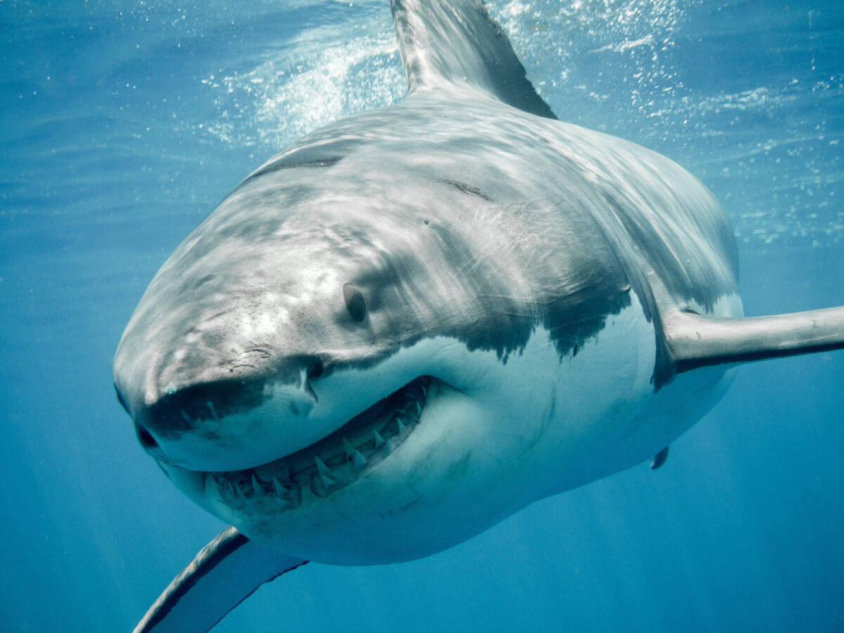
(Credit: © ramoncarretero - stock.adobe.com)
In a Nutshell
- Fast-swimming sharks like the shortfin mako and white shark carry their largest vertebrae in the middle of the body, with increasingly reinforced backbones toward the tail to power high-speed swimming.
- Common thresher sharks, which stun prey by whipping their long tails, have the highest number of internal reinforcing structures of any species studied, likely to handle extreme bending forces.
- As sharks grow larger, mineral builds up more slowly than the body lengthens. Rather than scaling up the size of each vertebra, adult sharks appear to reorganize their internal architecture, a result that ran counter to what the researchers expected.
Great white sharks, mako sharks, and their relatives may all look like sleek predators from the outside, but their backbones tell surprisingly different stories. A new study used high-powered 3D scanning to look inside the vertebrae of six shark species, revealing how evolution has shaped each animal’s skeleton to match the way it moves through the water.
Sharks have cartilage skeletons instead of bone, but that cartilage is partially hardened with calcium deposits in patterns that vary from species to species. Scientists at Florida Atlantic University and the National Oceanic and Atmospheric Administration, writing in the Journal of Anatomy, examined 139 vertebrae from 24 sharks across six species: shortfin mako, white shark, porbeagle, sand tiger, common thresher shark, and basking shark. Using a scanning method that works like an extremely detailed X-ray and can build three-dimensional images, they mapped the size, shape, and internal mineral structures of vertebrae from the front, middle, and back of each shark’s body.
What emerged was a detailed picture of skeletal engineering tuned to each animal’s lifestyle, from the blistering speed of a mako to the whip-cracking tail strike of a thresher shark.
How Shark Vertebrae Power High-Speed Swimming
Shortfin mako, porbeagle, and white sharks are fast, athletic predators built for speed. All three concentrate their swimming motion in the tail region, making them highly efficient at cruising long distances. Their skeletal architecture reflects this precisely.
In these species, the largest vertebrae sit in the mid-body, giving the torso stability while keeping the rear flexible enough for rapid tail movement. Moving toward the tail, the vertebrae get shorter and pack in more internal reinforcing plates, thin mineralized structures that radiate outward inside each vertebra like spokes. More of these plates are thought to stiffen the vertebrae, and stiffer vertebrae can pass energy from muscle to tail more efficiently. Earlier research on an artificial, shark-like vertebral column found that greater stiffness went hand in hand with faster swimming, a pattern the structural findings here appear to support.
While shortfin mako and white sharks showed very similar vertebral structures throughout, porbeagles stood apart from their close relatives in meaningful ways. Porbeagles had the lowest precaudal vertebral counts of the fast lamnid sharks examined, and their individual vertebrae have a different geometry. Researchers think this difference might reflect a structural workaround for having a shorter spinal column while still supporting the same fast-swimming style, though they stop short of confirming it as a mechanism.
A Tail Built Like a Weapon
Perhaps the most dramatic findings came from the common thresher shark, an animal famous for hunting by slapping prey with its elongated upper tail fin, sometimes in overhead whipping motions, sometimes sideways. This behavior puts enormous, multidirectional stress on the spine, and the skeleton appears to have evolved accordingly.
Common thresher sharks had the highest count of internal reinforcing plates of any species studied, averaging 29.1 per vertebra, along with the highest average number of branching points on those structures. Researchers believe the extra reinforcements help spread the stress of extreme bending across a wider area, much like the struts in a bridge. Their vertebrae are also flattened front-to-back compared to other species, a shape the study links to limiting excessive bending between individual vertebrae and giving the shark more controlled, precise tail movements during strikes. According to the authors, that front-to-back compression paired with high plate counts stands out as a signature of common thresher shark vertebrae, well suited to handling loads coming from several directions at once.

Slow Swimmers Have Their Own Skeletal Logic
Not every shark in the study is built for speed or dramatic hunting. Sand tigers and basking sharks swim slowly and deliberately, and their vertebrae reflect a different set of priorities.
Sand tigers had the fewest internal reinforcing plates of any species examined, which the researchers link to less stiff, more flexible vertebrae likely suited to the slow, maneuverable swimming they use in cluttered nearshore waters. Their vertebrae were largest at the front of the body and shrank moving toward the tail, the reverse of the pattern seen in fast-swimming species. Despite having few reinforcing plates, sand tigers had unexpectedly high numbers of branching points within those structures, which researchers think may help spread the smaller, routine stresses that come with slow maneuvering.
Basking sharks, filter feeders that cruise slowly near the surface, showed dramatically reduced hardening throughout their vertebrae. Because their vertebrae lacked well-defined internal structures, researchers left basking sharks out of the mineral analysis entirely rather than attempt counts that would not be reliable. The study could assess overall shape: basking sharks have unusually elongated, deeply concave vertebrae. The paper offers these features as a possible fit for slow swimming against the heavy drag created by open-mouth surface feeding, while treating the idea as a tentative interpretation rather than a settled conclusion.
Shark Vertebrae Rebuild From the Inside as Sharks Grow
One of the study’s more surprising results concerns how shark vertebrae change as the animals grow. Researchers expected larger sharks to simply have larger vertebrae with proportionally more internal structures. Instead, in adult sharks, mineral volume scaled more slowly than body length. Rather than depositing bulk that adds proportionally to each vertebra’s size, adult sharks appear to reorganize their internal architecture, an interpretation the authors say warrants further study.
Among common thresher sharks specifically, larger individuals had more branching points but not more reinforcing plates, indicating that as the body grows heavier and generates more force during tail strikes, the skeleton responds by building a denser internal support network rather than scaling everything up uniformly.
Compared with bone, mineralized cartilage in shark backbones has been studied far less, and the three-dimensional details of how it hardens are still poorly understood. This study begins to fill that gap. By combining 3D imaging and shape analysis across six species, the researchers showed that hardened cartilage in shark vertebrae is a finely engineered material, arranged to serve very specific mechanical purposes that differ not only between species but along the length of a single animal’s spine. Mapping that variation adds to a broader understanding of how mineralized cartilage supports movement and ecology. More immediately, it reframes how scientists picture the shark skeleton: not as a rough draft of a bony one, but as a precision instrument, built from the inside out.
Paper Notes
Limitations
This study was limited by sample size in certain species, particularly the basking shark, for which only one specimen was available, and the sand tiger, for which only two specimens were examined. Researchers acknowledge this restricts the conclusions that can be drawn for those species, and note that one sand tiger specimen came from a managed care facility, which may have influenced its skeletal development. The study also did not examine intervertebral joint length or vertebral counts in relation to body length, factors the authors acknowledge are likely crucial to understanding how the whole column works mechanically. Basking shark vertebrae were excluded from the mineral analysis because of their poor calcification, and no posterior vertebrae were available for the basking shark specimen. Regional sampling was not always complete across individuals, as some sharks did not have vertebrae available from all three body locations. The authors recommend future work using finite-element analysis, additional species from other shark orders, and larger sample sizes.
Funding and Disclosures
This work was supported by a National Science Foundation CAREER Grant (NSF, IOS-1941713) awarded to Marianne Porter. Additional support for Jamie L. Knaub came from the Jim Elliot Award from Tomography for Scientific Advancement (ToScA), along with the Vincent Saurino Fellowship, the Newell Doctoral Fellowship, the National Save the Sea Turtle Foundation Scholarship, the Graduate Fellowship for Academic Excellence, and the Rosalyn E. Schonzeit Scholarship from Florida Atlantic University. Lisa J. Natanson is noted as retired. This is an open-access article published under a Creative Commons Attribution License. U.S. Government employees contributed portions of the work, which are in the public domain in the USA.
Publication Details
Authors: Jamie L. Knaub, Madisan Biordi, Emma Pawlik, Michelle Passerotti, Lisa J. Natanson, Tricia Meredith, and Marianne Porter. Knaub, Biordi, Pawlik, and Porter are affiliated with the Department of Biological Sciences, Florida Atlantic University, Boca Raton, Florida. Passerotti and Natanson are affiliated with the Apex Predators Program, Northeast Fisheries Science Center, National Oceanic and Atmospheric Administration, Narragansett, Rhode Island. Meredith is affiliated with FAU Lab Schools, College of Education, Florida Atlantic University, Boca Raton, Florida. Journal: Journal of Anatomy
Year: 2026
Paper Title: “Skeletons of swiftly swimming sharks: Three-dimensional analysis of lamniform vertebral morphology and mineral architecture”
DOI: 10.1111/joa.70209


