
Visualization from the SHIC-XE AI framework revealing the anatomical cues used by AI to detect pain in horses. (Credit: Tel-Hai University of Kiryat Shmona in the Galilee)
In a Nutshell
- A new AI framework called SHIC-XE maps a horse pain-detection model’s attention onto a stable 3D horse model, addressing flickering, inconsistent visual explanations in video analysis.
- When the AI’s attention was compared with veterinary expert pain scores, meaningful correlations emerged for the ears and cheek muscles, but the eyes showed a negative correlation, meaning the AI and experts diverged on that cue.
- The study provides the first quantitative check of an AI pain-recognition explanation system against domain expert ratings in this context, pushing back on a common assumption that AI explanations which look plausible are automatically meaningful in a clinical setting.
Horses are masters of disguise when it comes to pain. As prey animals, they evolved to hide signs of suffering around potential threats, including the humans tasked with caring for them. That behavioral tendency has made pain assessment in horses notoriously difficult, pushing veterinarians to rely on trained observation of subtle facial changes. Now, a research team has built an AI explainability framework for horse pain-recognition models, one that maps where those models focus in a video onto a stable 3D model of a horse’s face and then measures that map against what real equine experts say matters. What they found both validates and complicates the growing field of AI-powered animal pain recognition.
Published in the International Journal of Computer Vision in 2026, the study introduces a system called SHIC-XE, designed to solve a persistent and underappreciated problem in AI explainability: when a camera records a moving animal, the AI’s visual attention maps tend to flicker and jump around frame by frame, making it nearly impossible to understand what the system is actually learning. SHIC-XE addresses this by projecting the AI’s attention onto a stable, three-dimensional model of a horse, so that no matter what angle or pose appears in the video, the system always anchors its observations to the same physical reference points on the face.
Perhaps more importantly, the researchers did something surprisingly rare in this field: they compared the AI’s attention patterns against pain scores assigned by trained veterinary clinicians, using an established tool called the Horse Grimace Scale. That comparison revealed meaningful, though modest, correlations in some facial regions and a puzzling negative correlation in others, raising pointed questions about whether AI systems that look plausible are actually reasoning the way experts do.
How Horses Hide Pain, and Why That Makes AI Hard to Trust
Veterinary pain assessment has long depended on human observation. For horses specifically, clinicians use a validated scoring system built around six distinct facial signals: ear position, tension above the eye area, eye tightening, cheek muscle strain, chin tension, and nostril strain. Each feature is scored on a simple scale, and a horse in significant pain tends to show specific, recognizable combinations of these signals, though detecting them requires training and careful attention.
Automating that process with AI is appealing because it removes variability across observers and allows continuous monitoring. But past AI systems built for equine pain recognition have struggled in a specific way: they might perform reasonably well at classifying pain versus no pain, yet offer explanations for their decisions that don’t hold up to scrutiny. A system might highlight a patch of background instead of a facial feature, or produce heat maps that shift wildly between frames of the same video. Clinicians looking at those explanations can’t reliably trust that the AI is reasoning about the right things.
SHIC-XE was built to address that trust problem directly. Rather than generating a fresh heat map for every individual video frame, the system learns dense connections between flat, two-dimensional images and a three-dimensional surface model of a horse’s face. Every pixel in every frame is mapped to a specific point on that 3D model, so attention across an entire video can be cleanly averaged at consistent locations. A highlight on the nose in frame one and a highlight on the nose in frame two actually add up, rather than getting lost in the noise.
Three Datasets, Three Kinds of Horse Pain
To test the system, the researchers used three separate datasets covering different types of horse pain. The first involved 39 horses recorded before and after routine castration surgery, capturing post-surgical discomfort. The second followed six horses with experimentally induced joint inflammation. The third involved 11 horses and used a device applied to a horse’s upper lip as an acute pain stimulus.
For video analysis, the researchers used leave-one-subject-out testing, meaning the model was evaluated on a horse kept out of its training data, which prevents the system from simply memorizing individual animals. Across the three datasets, video-level performance scores were 0.67, 0.80, and 0.70, respectively, on a standard balanced metric that accounts for both missed pain detections and false alarms. The authors say the approach achieves classification performance comparable to earlier methods while avoiding the need for separate AI models trained on each facial region.
One advantage the researchers point to is architectural simplicity. Earlier approaches to automated equine pain recognition often trained a different classifier for each facial area, creating long chains of processing steps where an error at any link could cascade and break the whole system. SHIC-XE uses a single classifier paired with the correspondence mapping, meaning the entire system only needs to successfully locate the horse’s head once before the 3D mapping takes over.
When the AI and the Experts Disagree on Horse Pain
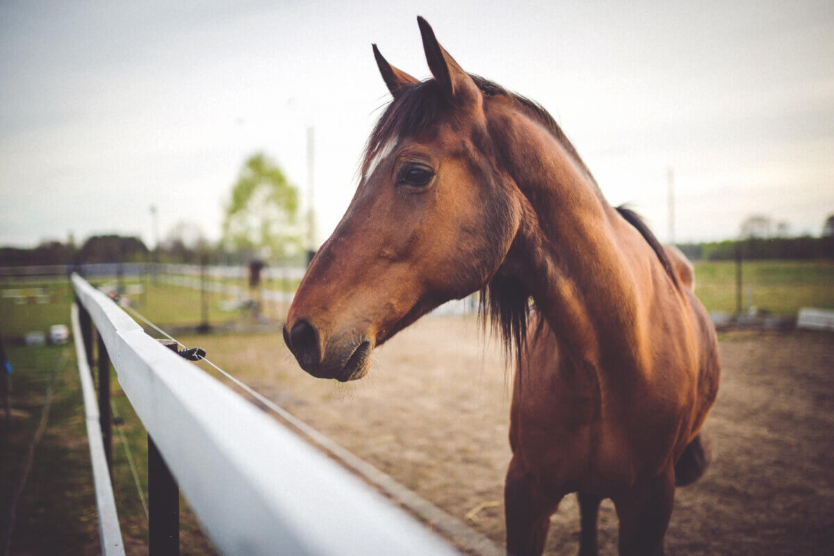
Comparison between machine attention and human expert scoring is the most revealing portion of the study. Using a refined set of 114 images from the first dataset, drawn from material that equine clinicians had scored with the Horse Grimace Scale, the researchers calculated whether the regions the AI paid most attention to correlated with the regions experts rated as most informative for pain.
For the ears, there was a meaningful positive correlation, meaning that when experts saw strong pain signals in ear position, the AI was also directing relatively more attention to that area. Cheek muscles showed a similar, though smaller, positive correlation. Both findings point to some genuine alignment between machine reasoning and veterinary expertise in those regions.
But the eyes told a different story. That region showed a statistically significant negative correlation, meaning the AI tended to look away from the eyes precisely when experts were rating eye-related pain signals as prominent. Mouth, nostril, and above-eye regions showed no meaningful correlations at all.
Researchers also found the AI concentrated notable attention on facial areas not directly featured in the Horse Grimace Scale, particularly the forehead and the bridge of the nose. Rather than treating this as a failure, the team offers an alternative interpretation: deep learning models may be detecting subtle tension in facial tissue that is real but too fine-grained for human observers to reliably score manually. Whether that marks a genuine discovery of complementary pain markers or simply reflects the AI picking up on patterns that don’t actually mean anything is a question the study cannot fully resolve.
A running theme throughout the paper is a pointed critique of how AI explainability gets evaluated in medical and veterinary settings. Most published studies in this space display appealing heat maps and invite experts to confirm they look reasonable, an approach the SHIC-XE team argues is not rigorous enough. By building measures that can be statistically tested against expert ratings, the researchers propose a higher standard for judging whether an AI is actually trustworthy.
According to the authors, the framework is not limited to horses. The underlying approach of mapping AI attention onto three-dimensional structural models could apply to any domain where spatial consistency between video footage and physical anatomy matters, from human medical imaging to quality inspection in manufacturing.
Still, the study’s own authors are clear that what they have achieved is preliminary validation, not definitive proof of clinical readiness. Small dataset sizes, particularly just six horses in the joint inflammation group, limit how confident anyone should be in the results. Manual selection of representative video frames for scoring may have inflated performance figures compared to what would be seen with randomly chosen frames. Errors in the 3D correspondence mapping, especially when a horse’s face is partially hidden or at an extreme angle, could distort the aggregated attention maps in ways that haven’t yet been fully characterized.
What the study establishes is a methodology: a way to take the subjective business of looking at AI attention visualizations and turn it into something that can actually be measured, tested, and challenged. In a field that has too often settled for visual plausibility as a proxy for trustworthiness, that methodological shift may matter as much as anything else the system can do.
Paper Notes
Limitations
Several important constraints shape how these findings should be read. Dataset sizes are small, with 39 horses in the post-surgical group, only six horses in the joint inflammation group, and 11 in the lip-twitch group, which limits statistical power and the ability to generalize results to broader horse populations. The researchers acknowledge that the image-based analysis relied on manually selected frames chosen for optimal quality, following the established Horse Grimace Scale protocol, but that this selection process may have produced inflated classification performance compared to randomly sampled frames. The framework depends on accurate three-dimensional correspondence mapping, and errors caused by occlusion or extreme head positions could distort the aggregated attention maps. Additionally, the system currently uses a single canonical 3D horse model, and breed-specific differences in head shape, such as between Arabian horses and draft breeds, could affect the spatial accuracy of explanations. The authors also distinguish between temporal stability, which their system achieves, and the analysis of how pain expressions evolve dynamically over time, noting that their mean aggregation approach compresses the time dimension and may miss transient micro-expressions. The paper also notes computational latency as a practical limitation.
Funding and Disclosures
Tel Hai Academic College provided open access funding. According to the paper, Marcelo Feighelstein, Annika Bremhorst, Claudia Spadavecchia, and Anna Zamansky were supported by an SNSF-ISF binational Switzerland-Israel project, with grant numbers Israel 1050/24 and Switzerland IC00I0L-228023. The authors declare no competing financial interests or personal relationships that could have influenced the work. All animal experiments were conducted under approved ethical protocols, with separate institutional approvals for each dataset as detailed in the paper.
Publication Details
Authors: Marcelo Feighelstein, Omer Bibi, Ofer Rozenbaum, Nathali Adrielli Agassi De Sales, Guilherme Camargo Ferraz, Ilan Shimshoni, Dirk van der Linden, Emanuela Dalla Costa, Annika Bremhorst, Claudia Spadavecchia, Anna Zamansky
Journal: International Journal of Computer Vision (2026), Volume 134, Article 342
Paper Title: “SHIC-XE: Viewpoint-Invariant Explainability via Dense 2D-3D Correspondences: an Application to Equine Pain Recognition”
DOI: 10.1007/s11263-026-02910-3
Published online: July 10, 2026


